 Movie
Movie Controller

[English] 日本語
 Yorodumi
- PDB-5fd0: Streptomyces plicatus N-acetyl-beta-hexosaminidase in complex wit... -
+ Open data

- Basic information
| Entry | Database: PDB / ID: 5fd0 | ||||||
|---|---|---|---|---|---|---|---|
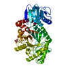
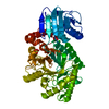
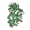
| Title | Streptomyces plicatus N-acetyl-beta-hexosaminidase in complex with NAGlucal | ||||||
 Components Components | B-N-acetylhexosaminidase | ||||||
 Keywords Keywords |  HYDROLASE / Family 20 glycoside hydrolase / SpHex / GH20 / NAG-glucal / hexosaminidase / glycosidase inhibitor HYDROLASE / Family 20 glycoside hydrolase / SpHex / GH20 / NAG-glucal / hexosaminidase / glycosidase inhibitor | ||||||
| Function / homology |  Function and homology information Function and homology informationglycosaminoglycan metabolic process / beta-N-acetylhexosaminidase / N-acetyl-beta-D-galactosaminidase activity / ganglioside catabolic process / beta-N-acetylglucosaminidase activity / lysosome / carbohydrate metabolic process / membraneSimilarity search - Function | ||||||
| Biological species |  Streptomyces plicatus (bacteria) Streptomyces plicatus (bacteria) | ||||||
| Method | X-RAY DIFFRACTION / MOLECULAR REPLACEMENT / Resolution: 2 Å | ||||||
 Authors Authors | Vadlamani, G. / Mark, B.L. | ||||||
| Funding support |  Canada, 1items Canada, 1items
| ||||||
 Citation Citation | Journal: Chem.Commun.(Camb.) / Year: 2016 Title: N-Acetyl glycals are tight-binding and environmentally insensitive inhibitors of hexosaminidases. Authors: Santana, A.G. / Vadlamani, G. / Mark, B.L. / Withers, S.G. | ||||||
| History |
|
- Structure visualization
| Structure viewer | Molecule: MolmilJmol/JSmol |
|---|
- Downloads & links
-Download
| PDBx/mmCIF format | 5fd0.cif.gz | 210.7 KB | Display | PDBx/mmCIF format |
|---|---|---|---|---|
| PDB format | pdb5fd0.ent.gz | 166.8 KB | Display | PDB format |
| PDBx/mmJSON format | 5fd0.json.gz | Tree view | PDBx/mmJSON format | |
| Others |  Other downloads Other downloads |
-Validation report
| Arichive directory | https://data.pdbj.org/pub/pdb/validation_reports/fd/5fd0ftp://data.pdbj.org/pub/pdb/validation_reports/fd/5fd0 | HTTPS FTP |
|---|
-Related structure data
| Related structure data | 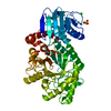 5fczC  1hp4S S: Starting model for refinement C: citing same article ( |
|---|---|
| Similar structure data |


-Links
 PDBj
PDBj
- Assembly
| Deposited unit | 
| |||||||||||||||||||||
|---|---|---|---|---|---|---|---|---|---|---|---|---|---|---|---|---|---|---|---|---|---|---|
| 1 |
| |||||||||||||||||||||
| 2 | 
| |||||||||||||||||||||
| Unit cell |
| |||||||||||||||||||||
| Components on special symmetry positions |
| |||||||||||||||||||||
| Details | monomer according to gel filtration |
-Components
| #1: Protein | Mass: 56126.777 Da / Num. of mol.: 1 Source method: isolated from a genetically manipulated source Source: (gene. exp.) Streptomyces plicatus (bacteria) / Gene: hex / Plasmid: pET-3a / Details (production host): Ampicillin resistance / Production host: Escherichia coli (E. coli) / Strain (production host): BL21-Gold(DE3) / References: UniProt: O85361 | ||||
|---|---|---|---|---|---|
| #2: Sugar | ChemComp-NAG / N-Acetylglucosamine  Type: D-saccharide, beta linking / Mass: 221.208 Da / Num. of mol.: 1 Type: D-saccharide, beta linking / Mass: 221.208 Da / Num. of mol.: 1Source method: isolated from a genetically manipulated source Formula: C8H15NO6 | ||||
| #3: Chemical | ChemComp-SO4 / Sulfate  Mass: 96.063 Da / Num. of mol.: 4 / Source method: obtained synthetically / Formula: SO4 Mass: 96.063 Da / Num. of mol.: 4 / Source method: obtained synthetically / Formula: SO4#4: Chemical | Glycerol  Mass: 92.094 Da / Num. of mol.: 2 / Source method: isolated from a natural source / Formula: C3H8O3 Mass: 92.094 Da / Num. of mol.: 2 / Source method: isolated from a natural source / Formula: C3H8O3#5: Water | ChemComp-HOH / | Water Mass: 18.015 Da / Num. of mol.: 677 / Source method: isolated from a natural source / Formula: H2O Mass: 18.015 Da / Num. of mol.: 677 / Source method: isolated from a natural source / Formula: H2O |
-Experimental details
-Experiment
| Experiment | Method: X-RAY DIFFRACTION |
|---|
- Sample preparation
| Crystal | Density Matthews: 4.03 Å3/Da / Density % sol: 69.44 % |
|---|---|
| Crystal grow | Temperature: 293.15 K / Method: vapor diffusion, hanging drop / pH: 6 Details: 1.8M ammonium sulphate, 0.1M trisodium citrate pH 6.0 |
-Data collection
| Diffraction | Mean temperature: 100 K |
|---|---|
| Diffraction source | Source: ROTATING ANODE / Type: RIGAKU MICROMAX-007 HF / Wavelength: 1.5418 Å |
| Detector | Type: RIGAKU RAXIS IV++ / Detector: IMAGE PLATE / Date: May 19, 2015 |
| Radiation | Protocol: SINGLE WAVELENGTH / Monochromatic (M) / Laue (L): M / Scattering type: x-ray |
| Radiation wavelength | Wavelength: 1.5418 Å / Relative weight: 1 |
| Reflection | Resolution: 2→54.81 Å / Num. obs: 61567 / % possible obs: 98.2 % / Redundancy: 7.2 % / Biso Wilson estimate: 23.06 Å2 / Rmerge(I) obs: 0.123 / Net I/σ(I): 11.1 |
| Reflection shell | Resolution: 2→2.05 Å / Redundancy: 6.6 % / Rmerge(I) obs: 0.704 / Mean I/σ(I) obs: 2.3 / % possible all: 94.1 |
- Processing
| Software |
| |||||||||||||||||||||||||||||||||||||||||||||||||||||||||||||||||||||||||||||||||||||||||||||||||||||||||
|---|---|---|---|---|---|---|---|---|---|---|---|---|---|---|---|---|---|---|---|---|---|---|---|---|---|---|---|---|---|---|---|---|---|---|---|---|---|---|---|---|---|---|---|---|---|---|---|---|---|---|---|---|---|---|---|---|---|---|---|---|---|---|---|---|---|---|---|---|---|---|---|---|---|---|---|---|---|---|---|---|---|---|---|---|---|---|---|---|---|---|---|---|---|---|---|---|---|---|---|---|---|---|---|---|---|---|
| Refinement | Method to determine structure: MOLECULAR REPLACEMENT Starting model: 1HP4 Resolution: 2→33.759 Å / SU ML: 0.19 / Cross valid method: FREE R-VALUE / σ(F): 1.34 / Phase error: 17.28 / Stereochemistry target values: ML
| |||||||||||||||||||||||||||||||||||||||||||||||||||||||||||||||||||||||||||||||||||||||||||||||||||||||||
| Solvent computation | Shrinkage radii: 0.9 Å / VDW probe radii: 1.11 Å / Solvent model: FLAT BULK SOLVENT MODEL | |||||||||||||||||||||||||||||||||||||||||||||||||||||||||||||||||||||||||||||||||||||||||||||||||||||||||
| Displacement parameters | Biso mean: 25.41 Å2 | |||||||||||||||||||||||||||||||||||||||||||||||||||||||||||||||||||||||||||||||||||||||||||||||||||||||||
| Refinement step | Cycle: LAST / Resolution: 2→33.759 Å
| |||||||||||||||||||||||||||||||||||||||||||||||||||||||||||||||||||||||||||||||||||||||||||||||||||||||||
| Refine LS restraints |
| |||||||||||||||||||||||||||||||||||||||||||||||||||||||||||||||||||||||||||||||||||||||||||||||||||||||||
| LS refinement shell |
|
