 Movie
Movie Controller

[English] 日本語
 Yorodumi
- PDB-5f7k: Blood group antigen binding adhesin BabA of Helicobacter pylori s... -
+ Open data

- Basic information
| Entry | Database: PDB / ID: 5f7k | ||||||||||||||||||
|---|---|---|---|---|---|---|---|---|---|---|---|---|---|---|---|---|---|---|---|
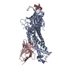
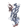
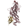
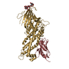
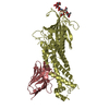
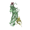
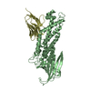
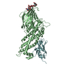
| Title | Blood group antigen binding adhesin BabA of Helicobacter pylori strain 17875 in complex with Nanobody Nb-ER19 | ||||||||||||||||||
 Components Components |
| ||||||||||||||||||
 Keywords Keywords |  CELL ADHESION / Adhesin / Lectin / Nanobody / Complex CELL ADHESION / Adhesin / Lectin / Nanobody / Complex | ||||||||||||||||||
| Function / homology |  Function and homology information Function and homology informationSabA, N-terminal extracellular adhesion domain / SabA N-terminal extracellular adhesion domain / Outer membrane protein, Helicobacter / Helicobacter outer membrane protein / Immunoglobulins / Immunoglobulin-like / Sandwich / Mainly BetaSimilarity search - Domain/homology | ||||||||||||||||||
| Biological species |   Helicobacter pylori (bacteria) Helicobacter pylori (bacteria) Lama glama (llama) Lama glama (llama) | ||||||||||||||||||
| Method | X-RAY DIFFRACTION / SYNCHROTRON / MOLECULAR REPLACEMENT / Resolution: 2.17 Å | ||||||||||||||||||
 Authors Authors | Moonens, K. / Gideonsson, P. / Subedi, S. / Romao, E. / Oscarson, S. / Muyldermans, S. / Boren, T. / Remaut, H. | ||||||||||||||||||
| Funding support |  Belgium, Belgium,  Sweden, 5items Sweden, 5items
| ||||||||||||||||||
 Citation Citation | Journal: Cell Host Microbe / Year: 2016 Title: Structural Insights into Polymorphic ABO Glycan Binding by Helicobacter pylori. Authors: Moonens, K. / Gideonsson, P. / Subedi, S. / Bugaytsova, J. / Romao, E. / Mendez, M. / Norden, J. / Fallah, M. / Rakhimova, L. / Shevtsova, A. / Lahmann, M. / Castaldo, G. / Brannstrom, K. / ...Authors: Moonens, K. / Gideonsson, P. / Subedi, S. / Bugaytsova, J. / Romao, E. / Mendez, M. / Norden, J. / Fallah, M. / Rakhimova, L. / Shevtsova, A. / Lahmann, M. / Castaldo, G. / Brannstrom, K. / Coppens, F. / Lo, A.W. / Ny, T. / Solnick, J.V. / Vandenbussche, G. / Oscarson, S. / Hammarstrom, L. / Arnqvist, A. / Berg, D.E. / Muyldermans, S. / Boren, T. / Remaut, H. | ||||||||||||||||||
| History |
|
- Structure visualization
| Structure viewer | Molecule: MolmilJmol/JSmol |
|---|
- Downloads & links
-Download
| PDBx/mmCIF format | 5f7k.cif.gz | 419.3 KB | Display | PDBx/mmCIF format |
|---|---|---|---|---|
| PDB format | pdb5f7k.ent.gz | 352.9 KB | Display | PDB format |
| PDBx/mmJSON format | 5f7k.json.gz | Tree view | PDBx/mmJSON format | |
| Others |  Other downloads Other downloads |
-Validation report
| Arichive directory | https://data.pdbj.org/pub/pdb/validation_reports/f7/5f7kftp://data.pdbj.org/pub/pdb/validation_reports/f7/5f7k | HTTPS FTP |
|---|
-Related structure data
| Related structure data | 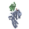 5f7lC 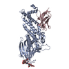 5f7mC  5f7nC 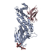 5f7wC  5f7yC 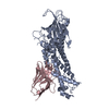 5f8qC 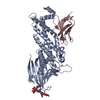 5f8rC 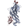 5f93C 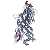 5f97C 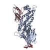 5f9aC 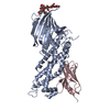 5f9dC C: citing same article ( |
|---|---|
| Similar structure data |


-Links
 PDBj
PDBj


- Assembly
| Deposited unit | 
| ||||||||||||||||||||||||||||||||||||||||||||||||
|---|---|---|---|---|---|---|---|---|---|---|---|---|---|---|---|---|---|---|---|---|---|---|---|---|---|---|---|---|---|---|---|---|---|---|---|---|---|---|---|---|---|---|---|---|---|---|---|---|---|
| 1 | 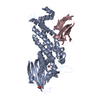
| ||||||||||||||||||||||||||||||||||||||||||||||||
| 2 | 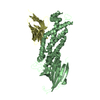
| ||||||||||||||||||||||||||||||||||||||||||||||||
| Unit cell |
| ||||||||||||||||||||||||||||||||||||||||||||||||
| Noncrystallographic symmetry (NCS) | NCS domain:
NCS domain segments:
NCS ensembles :
|
-Components
-Protein / Antibody , 2 types, 4 molecules ABCD
| #1: Protein | Mass: 49369.195 Da / Num. of mol.: 2 Source method: isolated from a genetically manipulated source Source: (gene. exp.) Helicobacter pylori (bacteria) / Gene: babA2 / Variant: 17875 / Production host: Escherichia coli (E. coli) / Variant (production host): Top10 / References: UniProt: O52269#2: Antibody | Mass: 13260.856 Da / Num. of mol.: 2 Source method: isolated from a genetically manipulated source Source: (gene. exp.) Lama glama (llama) / Production host: Escherichia coli (E. coli) / Variant (production host): WK6 |
|---|
-Non-polymers , 4 types, 484 molecules 


| #3: Chemical | ChemComp-PO4 / Phosphate Mass: 94.971 Da / Num. of mol.: 1 / Source method: obtained synthetically / Formula: PO4 Mass: 94.971 Da / Num. of mol.: 1 / Source method: obtained synthetically / Formula: PO4 |
|---|---|
| #4: Chemical | ChemComp-EDO / Ethylene glycol Mass: 62.068 Da / Num. of mol.: 1 / Source method: isolated from a natural source / Formula: C2H6O2 / Source: (gene. exp.) Helicobacter pylori (bacteria) / Gene: babA2 / Variant: 17875 / Production host: Escherichia coli (E. coli) / Variant (production host): Top10 Mass: 62.068 Da / Num. of mol.: 1 / Source method: isolated from a natural source / Formula: C2H6O2 / Source: (gene. exp.) Helicobacter pylori (bacteria) / Gene: babA2 / Variant: 17875 / Production host: Escherichia coli (E. coli) / Variant (production host): Top10 |
| #5: Chemical | ChemComp-NA /  Mass: 22.990 Da / Num. of mol.: 1 / Source method: obtained synthetically / Formula: Na Mass: 22.990 Da / Num. of mol.: 1 / Source method: obtained synthetically / Formula: Na |
| #6: Water | ChemComp-HOH / WaterMass: 18.015 Da / Num. of mol.: 481 / Source method: isolated from a natural source / Formula: H2O |
-Experimental details
-Experiment
| Experiment | Method: X-RAY DIFFRACTION |
|---|
- Sample preparation
| Crystal | Density Matthews: 3.42 Å3/Da / Density % sol: 64 % |
|---|---|
| Crystal grow | Temperature: 293 K / Method: vapor diffusion, sitting drop / pH: 6.5 Details: 0.2M sodium nitrate, 0.1M Bis Tris propane pH 6.5, 20% w/v PEG 3350 |
-Data collection
| Diffraction | Mean temperature: 100 K |
|---|---|
| Diffraction source | Source: SYNCHROTRON / Site: Diamond  / Beamline: I04 / Wavelength: 0.97949 Å / Beamline: I04 / Wavelength: 0.97949 Å |
| Detector | Type: DECTRIS PILATUS 6M-F / Detector: PIXEL / Date: Feb 5, 2014 |
| Radiation | Protocol: SINGLE WAVELENGTH / Monochromatic (M) / Laue (L): M / Scattering type: x-ray |
| Radiation wavelength | Wavelength: 0.97949 Å / Relative weight: 1 |
| Reflection | Resolution: 2.17→58.04 Å / Num. obs: 85215 / % possible obs: 99.4 % / Redundancy: 4.5 % / Biso Wilson estimate: 40.4 Å2 / Rmerge(I) obs: 0.046 / Net I/σ(I): 15.6 |
| Reflection shell | Resolution: 2.17→2.23 Å / Redundancy: 4.7 % / Rmerge(I) obs: 0.61 / Mean I/σ(I) obs: 2.2 / % possible all: 100 |
- Processing
| Software |
| ||||||||||||||||||||||||||||||||||||||||||||||||||||||||||||||||||||||||||||||||||||||||||||||||||||||||||||||||||||||||||||||||||||||||||||||||||||||||||||||||||||||||||||||||||||||
|---|---|---|---|---|---|---|---|---|---|---|---|---|---|---|---|---|---|---|---|---|---|---|---|---|---|---|---|---|---|---|---|---|---|---|---|---|---|---|---|---|---|---|---|---|---|---|---|---|---|---|---|---|---|---|---|---|---|---|---|---|---|---|---|---|---|---|---|---|---|---|---|---|---|---|---|---|---|---|---|---|---|---|---|---|---|---|---|---|---|---|---|---|---|---|---|---|---|---|---|---|---|---|---|---|---|---|---|---|---|---|---|---|---|---|---|---|---|---|---|---|---|---|---|---|---|---|---|---|---|---|---|---|---|---|---|---|---|---|---|---|---|---|---|---|---|---|---|---|---|---|---|---|---|---|---|---|---|---|---|---|---|---|---|---|---|---|---|---|---|---|---|---|---|---|---|---|---|---|---|---|---|---|---|
| Refinement | Method to determine structure: MOLECULAR REPLACEMENT / Resolution: 2.17→58.04 Å / Cor.coef. Fo:Fc: 0.967 / Cor.coef. Fo:Fc free: 0.954 / SU B: 8.463 / SU ML: 0.109 / Cross valid method: THROUGHOUT / ESU R: 0.161 / ESU R Free: 0.148 / Stereochemistry target values: MAXIMUM LIKELIHOOD / Details: HYDROGENS HAVE BEEN ADDED IN THE RIDING POSITIONS
| ||||||||||||||||||||||||||||||||||||||||||||||||||||||||||||||||||||||||||||||||||||||||||||||||||||||||||||||||||||||||||||||||||||||||||||||||||||||||||||||||||||||||||||||||||||||
| Solvent computation | Ion probe radii: 0.8 Å / Shrinkage radii: 0.8 Å / VDW probe radii: 1.2 Å / Solvent model: MASK | ||||||||||||||||||||||||||||||||||||||||||||||||||||||||||||||||||||||||||||||||||||||||||||||||||||||||||||||||||||||||||||||||||||||||||||||||||||||||||||||||||||||||||||||||||||||
| Displacement parameters | Biso mean: 54.385 Å2
| ||||||||||||||||||||||||||||||||||||||||||||||||||||||||||||||||||||||||||||||||||||||||||||||||||||||||||||||||||||||||||||||||||||||||||||||||||||||||||||||||||||||||||||||||||||||
| Refinement step | Cycle: LAST / Resolution: 2.17→58.04 Å
| ||||||||||||||||||||||||||||||||||||||||||||||||||||||||||||||||||||||||||||||||||||||||||||||||||||||||||||||||||||||||||||||||||||||||||||||||||||||||||||||||||||||||||||||||||||||
| Refine LS restraints |
|