 Movie
Movie Controller

[English] 日本語
 Yorodumi
- PDB-5f6i: Crystal Structure of Tier 2 Neutralizing Antibody DH428 from a Rh... -
+ Open data

- Basic information
| Entry | Database: PDB / ID: 5f6i | ||||||
|---|---|---|---|---|---|---|---|
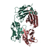
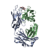
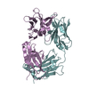
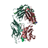
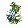
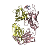
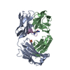
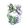
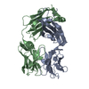
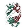
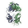
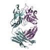
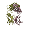
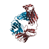
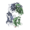
| Title | Crystal Structure of Tier 2 Neutralizing Antibody DH428 from a Rhesus Macaque | ||||||
 Components Components |
| ||||||
 Keywords Keywords |  IMMUNE SYSTEM / FAB FRAGMENT / HIV-1 / ANTIBODY IMMUNE SYSTEM / FAB FRAGMENT / HIV-1 / ANTIBODY | ||||||
| Function / homology | Immunoglobulins / Immunoglobulin-like / Sandwich / Mainly Beta Function and homology information Function and homology information | ||||||
| Biological species |  Macaca mulatta (Rhesus monkey) Macaca mulatta (Rhesus monkey) | ||||||
| Method | X-RAY DIFFRACTION / SYNCHROTRON / MOLECULAR REPLACEMENT / Resolution: 2.32 Å | ||||||
 Authors Authors | Fera, D. / Harrison, S.C. | ||||||
 Citation Citation | Journal: Cell Rep / Year: 2016 Title: Structural Constraints of Vaccine-Induced Tier-2 Autologous HIV Neutralizing Antibodies Targeting the Receptor-Binding Site. Authors: Bradley, T. / Fera, D. / Bhiman, J. / Eslamizar, L. / Lu, X. / Anasti, K. / Zhang, R. / Sutherland, L.L. / Scearce, R.M. / Bowman, C.M. / Stolarchuk, C. / Lloyd, K.E. / Parks, R. / Eaton, A. ...Authors: Bradley, T. / Fera, D. / Bhiman, J. / Eslamizar, L. / Lu, X. / Anasti, K. / Zhang, R. / Sutherland, L.L. / Scearce, R.M. / Bowman, C.M. / Stolarchuk, C. / Lloyd, K.E. / Parks, R. / Eaton, A. / Foulger, A. / Nie, X. / Karim, S.S. / Barnett, S. / Kelsoe, G. / Kepler, T.B. / Alam, S.M. / Montefiori, D.C. / Moody, M.A. / Liao, H.X. / Morris, L. / Santra, S. / Harrison, S.C. / Haynes, B.F. | ||||||
| History |
|
- Structure visualization
| Structure viewer | Molecule: MolmilJmol/JSmol |
|---|
- Downloads & links
-Download
| PDBx/mmCIF format | 5f6i.cif.gz | 175.2 KB | Display | PDBx/mmCIF format |
|---|---|---|---|---|
| PDB format | pdb5f6i.ent.gz | 137.3 KB | Display | PDB format |
| PDBx/mmJSON format | 5f6i.json.gz | Tree view | PDBx/mmJSON format | |
| Others |  Other downloads Other downloads |
-Validation report
| Arichive directory | https://data.pdbj.org/pub/pdb/validation_reports/f6/5f6iftp://data.pdbj.org/pub/pdb/validation_reports/f6/5f6i | HTTPS FTP |
|---|
-Related structure data
| Related structure data |  5f6hC  5f6jC 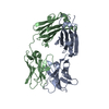 4qhlS C: citing same article ( S: Starting model for refinement |
|---|---|
| Similar structure data |


-Links
 PDBj
PDBj


- Assembly
| Deposited unit | 
| ||||||||
|---|---|---|---|---|---|---|---|---|---|
| 1 |
| ||||||||
| Unit cell |
|
-Components
| #1: Antibody | Mass: 24293.105 Da / Num. of mol.: 1 Source method: isolated from a genetically manipulated source Source: (gene. exp.) Macaca mulatta (Rhesus monkey) / Plasmid: pVRC-8400 / Cell line (production host): HEK 293T / Production host:  Homo sapiens (human) Homo sapiens (human) |
|---|---|
| #2: Antibody | Mass: 22926.225 Da / Num. of mol.: 1 Source method: isolated from a genetically manipulated source Source: (gene. exp.) Macaca mulatta (Rhesus monkey) / Plasmid: pVRC-8400 / Cell line (production host): HEK 293T / Production host: Homo sapiens (human) |
| #3: Water | ChemComp-HOH / Water Mass: 18.015 Da / Num. of mol.: 83 / Source method: isolated from a natural source / Formula: H2O Mass: 18.015 Da / Num. of mol.: 83 / Source method: isolated from a natural source / Formula: H2O |
-Experimental details
-Experiment
| Experiment | Method: X-RAY DIFFRACTION / Number of used crystals: 1 |
|---|
- Sample preparation
| Crystal | Density Matthews: 3.08 Å3/Da / Density % sol: 60.02 % |
|---|---|
| Crystal grow | Temperature: 293 K / Method: vapor diffusion, hanging drop / pH: 5 / Details: 40% PEG 400 and 100 mM sodium citrate, pH 5.0 |
-Data collection
| Diffraction | Mean temperature: 100 K |
|---|---|
| Diffraction source | Source: SYNCHROTRON / Site: APS  / Beamline: 24-ID-C / Wavelength: 0.97916 Å / Beamline: 24-ID-C / Wavelength: 0.97916 Å |
| Detector | Type: DECTRIS PILATUS 6M-F / Detector: PIXEL / Date: Feb 15, 2015 |
| Radiation | Protocol: SINGLE WAVELENGTH / Monochromatic (M) / Laue (L): M / Scattering type: x-ray |
| Radiation wavelength | Wavelength: 0.97916 Å / Relative weight: 1 |
| Reflection | Resolution: 2.32→60.36 Å / Num. obs: 22905 / % possible obs: 91.72 % / Redundancy: 4 % / Rmerge(I) obs: 0.086 / Net I/σ(I): 13.43 |
| Reflection shell | Resolution: 2.32→2.4 Å / Redundancy: 3.9 % / Rmerge(I) obs: 0.744 / Mean I/σ(I) obs: 2.2 / % possible all: 95 |
- Processing
| Software |
| ||||||||||||||||||||||||||||||||||||||||||||||||||||||||||||||||||||||
|---|---|---|---|---|---|---|---|---|---|---|---|---|---|---|---|---|---|---|---|---|---|---|---|---|---|---|---|---|---|---|---|---|---|---|---|---|---|---|---|---|---|---|---|---|---|---|---|---|---|---|---|---|---|---|---|---|---|---|---|---|---|---|---|---|---|---|---|---|---|---|---|
| Refinement | Method to determine structure: MOLECULAR REPLACEMENT Starting model: separated Fv and Fc regions of I3.2 Fab from PDB Entry 4QHL Resolution: 2.32→60.356 Å / FOM work R set: 0.8008 / SU ML: 0.3 / Cross valid method: FREE R-VALUE / σ(F): 1.35 / Phase error: 26.38 / Stereochemistry target values: ML
| ||||||||||||||||||||||||||||||||||||||||||||||||||||||||||||||||||||||
| Solvent computation | Shrinkage radii: 0.9 Å / VDW probe radii: 1.11 Å / Solvent model: FLAT BULK SOLVENT MODEL | ||||||||||||||||||||||||||||||||||||||||||||||||||||||||||||||||||||||
| Displacement parameters | Biso max: 106.72 Å2 / Biso mean: 48.6 Å2 / Biso min: 23.45 Å2 | ||||||||||||||||||||||||||||||||||||||||||||||||||||||||||||||||||||||
| Refinement step | Cycle: final / Resolution: 2.32→60.356 Å
| ||||||||||||||||||||||||||||||||||||||||||||||||||||||||||||||||||||||
| Refine LS restraints |
| ||||||||||||||||||||||||||||||||||||||||||||||||||||||||||||||||||||||
| LS refinement shell | Refine-ID: X-RAY DIFFRACTION / Total num. of bins used: 9
| ||||||||||||||||||||||||||||||||||||||||||||||||||||||||||||||||||||||
| Refinement TLS params. | Method: refined / Origin x: 3.0516 Å / Origin y: -18.8539 Å / Origin z: -21.5289 Å
| ||||||||||||||||||||||||||||||||||||||||||||||||||||||||||||||||||||||
| Refinement TLS group |
|
