- PDB-5f1z: Structure of TYK2 with inhibitor 16: 3-azanyl-5-[(2~{S})-3-methyl... -
+
Open data
ID or keywords:
Loading...
-
Basic information
Entry
Database: PDB / ID: 5f1z
Title
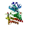
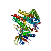
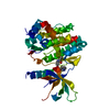
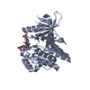
Structure of TYK2 with inhibitor 16: 3-azanyl-5-[(2~{S})-3-methylbutan-2-yl]-7-[1-methyl-5-(2-oxidanylpropan-2-yl)pyrazol-3-yl]-1~{H}-pyrazolo[4,3-c]pyridin-4-one
Protocol: SINGLE WAVELENGTH / Monochromatic (M) / Laue (L): M / Scattering type: x-ray
Radiation wavelength
Wavelength: 0.98 Å / Relative weight: 1
Reflection
Redundancy: 6.3 % / Number: 92057 / Rmerge(I) obs: 0.127 / Χ2: 1.03 / D res high: 2.35 Å / D res low: 50 Å / Num. obs: 14704 / % possible obs: 99
Diffraction reflection shell
Highest resolution (Å)
Lowest resolution (Å)
ID
Rmerge(I) obs
Chi squared
Redundancy
6.37
50
1
0.068
0.994
6
5.06
6.37
1
0.097
1.024
6.9
4.42
5.06
1
0.082
0.993
6.7
4.02
4.42
1
0.077
0.986
6.5
3.73
4.02
1
0.079
0.998
6.4
3.51
3.73
1
0.088
0.996
6.7
3.33
3.51
1
0.1
1.012
6.6
3.19
3.33
1
0.154
1.063
6.9
3.07
3.19
1
0.167
1.029
7
2.96
3.07
1
0.191
1.064
7.1
2.87
2.96
1
0.238
1.136
7.1
2.79
2.87
1
0.292
1.075
7.3
2.71
2.79
1
0.334
1.071
7.4
2.65
2.71
1
0.35
1.099
7
2.59
2.65
1
0.448
1.13
6
2.53
2.59
1
0.444
1.061
5.2
2.48
2.53
1
0.441
0.972
4.9
2.43
2.48
1
0.481
0.937
4.8
2.39
2.43
1
0.507
0.87
4
2.35
2.39
1
0.551
0.904
4
Reflection
Resolution: 2.35→50 Å / Num. obs: 14704 / % possible obs: 99 % / Redundancy: 6.3 % / Rmerge(I) obs: 0.127 / Χ2: 1.03 / Net I/av σ(I): 10.951 / Net I/σ(I): 7.1 / Num. measured all: 92057
Reflection shell
Diffraction-ID: 1 / Rejects: 0
Resolution (Å)
Redundancy (%)
Rmerge(I) obs
Num. unique all
Χ2
% possible all
2.35-2.39
4
0.551
660
0.904
94.6
2.39-2.43
4
0.507
665
0.87
95.1
2.43-2.48
4.8
0.481
681
0.937
96.2
2.48-2.53
4.9
0.441
682
0.972
97.6
2.53-2.59
5.2
0.444
707
1.061
99.4
2.59-2.65
6
0.448
737
1.13
98.8
2.65-2.71
7
0.35
669
1.099
99.4
2.71-2.79
7.4
0.334
741
1.071
99.7
2.79-2.87
7.3
0.292
690
1.075
100
2.87-2.96
7.1
0.238
761
1.136
100
2.96-3.07
7.1
0.191
697
1.064
99.9
3.07-3.19
7
0.167
741
1.029
99.9
3.19-3.33
6.9
0.154
715
1.063
100
3.33-3.51
6.6
0.1
745
1.012
99.7
3.51-3.73
6.7
0.088
743
0.996
99.6
3.73-4.02
6.4
0.079
752
0.998
99.7
4.02-4.42
6.5
0.077
755
0.986
100
4.42-5.06
6.7
0.082
789
0.993
100
5.06-6.37
6.9
0.097
815
1.024
100
6.37-50
6
0.068
959
0.994
99.8
-
Phasing
Phasing
Method: molecular replacement
Phasing MR
Model details: Phaser MODE: MR_AUTO
Highest resolution
Lowest resolution
Rotation
6.49 Å
41.24 Å
Translation
6.49 Å
41.24 Å
-
Processing
Software
Name
Version
Classification
HKL-2000
datacollection
SCALEPACK
datascaling
PHASER
2.5.1
phasing
REFMAC
refinement
PDB_EXTRACT
3.15
dataextraction
Refinement
Method to determine structure: MOLECULAR REPLACEMENT / Resolution: 2.65→30 Å / Cor.coef. Fo:Fc: 0.924 / Cor.coef. Fo:Fc free: 0.818 / WRfactor Rfree: 0.31 / WRfactor Rwork: 0.216 / FOM work R set: 0.7922 / SU B: 29.827 / SU ML: 0.276 / SU R Cruickshank DPI: 2.9907 / SU Rfree: 0.416 / Cross valid method: THROUGHOUT / σ(F): 0 / ESU R: 2.991 / ESU R Free: 0.416 / Stereochemistry target values: MAXIMUM LIKELIHOOD Details: HYDROGENS HAVE BEEN ADDED IN THE RIDING POSITIONS U VALUES : WITH TLS ADDED
Rfactor
Num. reflection
% reflection
Selection details
Rfree
0.31
514
4.9 %
RANDOM
Rwork
0.216
-
-
-
obs
0.2201
10006
99.8 %
-
Solvent computation
Ion probe radii: 0.8 Å / Shrinkage radii: 0.8 Å / VDW probe radii: 1.2 Å / Solvent model: BABINET MODEL WITH MASK
In the structure databanks used in Yorodumi, some data are registered as the other names, "COVID-19 virus" and "2019-nCoV". Here are the details of the virus and the list of structure data.
Jan 31, 2019. EMDB accession codes are about to change! (news from PDBe EMDB page)
EMDB accession codes are about to change! (news from PDBe EMDB page)
The allocation of 4 digits for EMDB accession codes will soon come to an end. Whilst these codes will remain in use, new EMDB accession codes will include an additional digit and will expand incrementally as the available range of codes is exhausted. The current 4-digit format prefixed with “EMD-” (i.e. EMD-XXXX) will advance to a 5-digit format (i.e. EMD-XXXXX), and so on. It is currently estimated that the 4-digit codes will be depleted around Spring 2019, at which point the 5-digit format will come into force.
The EM Navigator/Yorodumi systems omit the EMD- prefix.
Related info.:Q: What is EMD? / ID/Accession-code notation in Yorodumi/EM Navigator
Yorodumi is a browser for structure data from EMDB, PDB, SASBDB, etc.
This page is also the successor to EM Navigator detail page, and also detail information page/front-end page for Omokage search.
The word "yorodu" (or yorozu) is an old Japanese word meaning "ten thousand". "mi" (miru) is to see.
Related info.:EMDB / PDB / SASBDB / Comparison of 3 databanks / Yorodumi Search / Aug 31, 2016. New EM Navigator & Yorodumi / Yorodumi Papers / Jmol/JSmol / Function and homology information / Changes in new EM Navigator and Yorodumi
 Movie
Movie Controller

 Yorodumi
 Open data
 Basic information
 Components
 Keywords
 Kinase /
  Function and homology information
 Authors
 Citation
 Structure visualization
 Downloads & links
 Other downloads
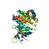


 PDBj
PDBj


 Assembly
 unidentified baculovirus
 Mass: 358.438 Da / Num. of mol.: 1 / Source method: obtained synthetically / Formula: C18H26N6O2
 Mass: 18.015 Da / Num. of mol.: 155 / Source method: isolated from a natural source / Formula: H2O
 Sample preparation
 / Beamline: 5.0.3 / Wavelength: 0.98 Å
 Processing