 Movie
Movie Controller

[English] 日本語
 Yorodumi
- PDB-5f1m: Crystal structure of Serine/threonine phosphatase Stp1 from Staph... -
+ Open data

- Basic information
| Entry | Database: PDB / ID: 5f1m | |||||||||
|---|---|---|---|---|---|---|---|---|---|---|
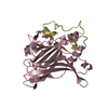
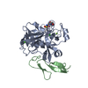
| Title | Crystal structure of Serine/threonine phosphatase Stp1 from Staphylococcus aureus | |||||||||
 Components Components | Phosphorylated protein phosphatase Phosphorylation Phosphorylation | |||||||||
 Keywords Keywords | HYDROLASE / Serine/threonine phosphatase 1 | |||||||||
| Function / homology |  Function and homology information Function and homology informationmyosin phosphatase activity / protein-serine/threonine phosphatase / metal ion bindingSimilarity search - Function | |||||||||
| Biological species |   Staphylococcus aureus (bacteria) Staphylococcus aureus (bacteria) | |||||||||
| Method | X-RAY DIFFRACTION / MOLECULAR REPLACEMENT / Resolution: 2.322 Å | |||||||||
 Authors Authors | Zheng, W.H. / Wang, T. / Li, Z.G. | |||||||||
| Funding support |  China, 2items China, 2items
| |||||||||
 Citation Citation | Journal: Cell Chem Biol / Year: 2016 Title: Structure-Based Identification of a Potent Inhibitor Targeting Stp1-Mediated Virulence Regulation in Staphylococcus aureus Authors: Zheng, W. / Cai, X. / Xie, M. / Liang, Y. / Wang, T. / Li, Z. | |||||||||
| History |
|
- Structure visualization
| Structure viewer | Molecule: MolmilJmol/JSmol |
|---|
- Downloads & links
-Download
| PDBx/mmCIF format | 5f1m.cif.gz | 120.3 KB | Display | PDBx/mmCIF format |
|---|---|---|---|---|
| PDB format | pdb5f1m.ent.gz | 92.8 KB | Display | PDB format |
| PDBx/mmJSON format | 5f1m.json.gz | Tree view | PDBx/mmJSON format | |
| Others |  Other downloads Other downloads |
-Validation report
| Arichive directory | https://data.pdbj.org/pub/pdb/validation_reports/f1/5f1mftp://data.pdbj.org/pub/pdb/validation_reports/f1/5f1m | HTTPS FTP |
|---|
-Related structure data
| Related structure data |  2pk0S S: Starting model for refinement |
|---|---|
| Similar structure data |


-Links
 PDBj
PDBj- Assembly
| Deposited unit | 
| ||||||||
|---|---|---|---|---|---|---|---|---|---|
| 1 |
| ||||||||
| Unit cell |
|
-Components
| #1: Protein | Phosphorylation / Protein phosphatase / Protein serine/threonine phosphatase PrpC%2C regulation of stationary phase Mass: 30646.371 Da / Num. of mol.: 1 Source method: isolated from a genetically manipulated source Source: (gene. exp.) Staphylococcus aureus (bacteria)Gene: prpC, AS858_01330, BN1321_240063, EQ80_005655, ER12_005650, ERS093009_00664 Plasmid: pCold I / Production host: Escherichia coli (E. coli) / Strain (production host): BL21 DE3 plysSReferences: UniProt: Q9RL81, protein-serine/threonine phosphatase | ||
|---|---|---|---|
| #2: Chemical | ChemComp-MN /   Mass: 54.938 Da / Num. of mol.: 4 / Source method: obtained synthetically / Formula: Mn Mass: 54.938 Da / Num. of mol.: 4 / Source method: obtained synthetically / Formula: Mn#3: Water | ChemComp-HOH / | Water Mass: 18.015 Da / Num. of mol.: 136 / Source method: isolated from a natural source / Formula: H2O Mass: 18.015 Da / Num. of mol.: 136 / Source method: isolated from a natural source / Formula: H2O |
-Experimental details
-Experiment
| Experiment | Method: X-RAY DIFFRACTION / Number of used crystals: 1 |
|---|
- Sample preparation
| Crystal | Density Matthews: 2.19 Å3/Da / Density % sol: 43.9 % |
|---|---|
| Crystal grow | Temperature: 293 K / Method: vapor diffusion, sitting drop / pH: 8.5 / Details: 15%PEG4000, 200mM MgCl2, 100mM Tris, 0.5mM MnCl2 / PH range: 8.2-8.7 |
-Data collection
| Diffraction | Mean temperature: 100 K | ||||||||||||||||||||||||||||||
|---|---|---|---|---|---|---|---|---|---|---|---|---|---|---|---|---|---|---|---|---|---|---|---|---|---|---|---|---|---|---|---|
| Diffraction source | Source: SEALED TUBE / Type: RIGAKU MICROMAX-002+ / Wavelength: 1.5418 Å | ||||||||||||||||||||||||||||||
| Detector | Type: RIGAKU SATURN 944+ / Detector: CCD / Date: Dec 31, 2014 | ||||||||||||||||||||||||||||||
| Radiation | Protocol: SINGLE WAVELENGTH / Monochromatic (M) / Laue (L): M / Scattering type: x-ray | ||||||||||||||||||||||||||||||
| Radiation wavelength | Wavelength: 1.5418 Å / Relative weight: 1 | ||||||||||||||||||||||||||||||
| Reflection | Resolution: 2.32→38.77 Å / Num. obs: 11662 / % possible obs: 100 % / Redundancy: 6.9 % / Biso Wilson estimate: 35.22 Å2 / CC1/2: 0.994 / Rmerge(I) obs: 0.177 / Rpim(I) all: 0.072 / Rrim(I) all: 0.192 / Net I/σ(I): 8.2 / Num. measured all: 80708 / Scaling rejects: 1 | ||||||||||||||||||||||||||||||
| Reflection shell | Diffraction-ID: 1 / Rejects: 0
|
- Processing
| Software |
| ||||||||||||||||||||||||||||||||||||||||
|---|---|---|---|---|---|---|---|---|---|---|---|---|---|---|---|---|---|---|---|---|---|---|---|---|---|---|---|---|---|---|---|---|---|---|---|---|---|---|---|---|---|
| Refinement | Method to determine structure: MOLECULAR REPLACEMENT Starting model: 2PK0 Resolution: 2.322→32.114 Å / SU ML: 0.3 / Cross valid method: NONE / σ(F): 1.34 / Phase error: 26.37 / Stereochemistry target values: ML
| ||||||||||||||||||||||||||||||||||||||||
| Solvent computation | Shrinkage radii: 0.9 Å / VDW probe radii: 1.11 Å / Solvent model: FLAT BULK SOLVENT MODEL | ||||||||||||||||||||||||||||||||||||||||
| Displacement parameters | Biso max: 123.18 Å2 / Biso mean: 43.9101 Å2 / Biso min: 17.25 Å2 | ||||||||||||||||||||||||||||||||||||||||
| Refinement step | Cycle: final / Resolution: 2.322→32.114 Å
| ||||||||||||||||||||||||||||||||||||||||
| Refine LS restraints |
| ||||||||||||||||||||||||||||||||||||||||
| LS refinement shell | Refine-ID: X-RAY DIFFRACTION / Total num. of bins used: 4 / % reflection obs: 100 %
| ||||||||||||||||||||||||||||||||||||||||
| Refinement TLS params. | Method: refined / Origin x: 48.9257 Å / Origin y: 96.1253 Å / Origin z: 183.1176 Å
| ||||||||||||||||||||||||||||||||||||||||
| Refinement TLS group | Selection details: chain 'A' |
