Entry Database : PDB / ID : 5f0vTitle X-ray crystal structure of a thiolase from Escherichia coli at 1.8 A resolution Acetyl-CoA acetyltransferase Keywords / / / / Function / homology Function Domain/homology Component
/ / / / / / / / / / / / / / / / / / / / / / / / / / / / / Biological species Escherichia coli K-12 (bacteria)Method / / Resolution : 1.8 Å Authors Ithayaraja, M. / Neelanjana, J. / Wierenga, R. / Savithri, H.S. / Murthy, M.R.N. Funding support Organization Grant number Country DBT BT/IN/FINNISH/27/MRNM/2009
Journal : Acta Crystallogr.,Sect.F / Year : 2016Title : Crystal structure of a thiolase from Escherichia coli at 1.8 angstrom resolution.Authors : Ithayaraja, M. / Janardan, N. / Wierenga, R.K. / Savithri, H.S. / Murthy, M.R. History Deposition Nov 28, 2015 Deposition site / Processing site Revision 1.0 Jul 13, 2016 Provider / Type Revision 1.1 Dec 18, 2019 Group / Derived calculationsCategory / citation_author / pdbx_struct_oper_listItem _citation.pdbx_database_id_PubMed / _citation.title ... _citation.pdbx_database_id_PubMed / _citation.title / _citation_author.name / _pdbx_struct_oper_list.symmetry_operation Revision 1.2 Nov 8, 2023 Group / Database references / Refinement descriptionCategory chem_comp_atom / chem_comp_bond ... chem_comp_atom / chem_comp_bond / database_2 / pdbx_initial_refinement_model / struct_ncs_dom_lim Item _database_2.pdbx_DOI / _database_2.pdbx_database_accession ... _database_2.pdbx_DOI / _database_2.pdbx_database_accession / _struct_ncs_dom_lim.beg_auth_comp_id / _struct_ncs_dom_lim.beg_label_asym_id / _struct_ncs_dom_lim.beg_label_comp_id / _struct_ncs_dom_lim.beg_label_seq_id / _struct_ncs_dom_lim.end_auth_comp_id / _struct_ncs_dom_lim.end_label_asym_id / _struct_ncs_dom_lim.end_label_comp_id / _struct_ncs_dom_lim.end_label_seq_id
Show all Show less
 Movie
Movie Controller

 Yorodumi
 Open data
 Basic information
 Components
 Keywords
 TRANSFERASE / E.coli thiolase /
  Function and homology information
 Authors
 India, 1items
  Citation
 Structure visualization
 Downloads & links
 Other downloads


 PDBj
PDBj

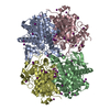
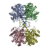
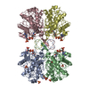
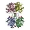
 Assembly

 Mass: 62.068 Da / Num. of mol.: 44 / Source method: obtained synthetically / Formula: C2H6O2
 Mass: 18.015 Da / Num. of mol.: 1136 / Source method: isolated from a natural source / Formula: H2O
 Sample preparation
 Processing