 Movie
Movie Controller


+ Open data

- Basic information
| Entry | Database: PDB / ID: 5erq | |||||||||
|---|---|---|---|---|---|---|---|---|---|---|
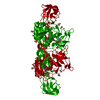
| Title | Gephyrin E domain at 1.55 angstrom resolution | |||||||||
 Components Components | Gephyrin | |||||||||
 Keywords Keywords | TRANSFERASE / Molybdenum cofactor / tungsten cofactor / Moco biosynthesis | |||||||||
| Function / homology |  Function and homology information Function and homology informationMolybdenum cofactor biosynthesis / molybdopterin cofactor biosynthetic process / glycine receptor clustering / molybdopterin adenylyltransferase / molybdopterin adenylyltransferase activity / establishment of synaptic specificity at neuromuscular junction / molybdopterin molybdotransferase / molybdopterin molybdotransferase activity / postsynaptic specialization / gamma-aminobutyric acid receptor clustering ...Molybdenum cofactor biosynthesis / molybdopterin cofactor biosynthetic process / glycine receptor clustering / molybdopterin adenylyltransferase / molybdopterin adenylyltransferase activity / establishment of synaptic specificity at neuromuscular junction / molybdopterin molybdotransferase / molybdopterin molybdotransferase activity / postsynaptic specialization / gamma-aminobutyric acid receptor clustering / nitrate reductase activity / inhibitory synapse / Mo-molybdopterin cofactor biosynthetic process / glycinergic synapse / postsynaptic neurotransmitter receptor diffusion trapping / postsynaptic specialization membrane / response to metal ion / molybdopterin cofactor binding / neurotransmitter receptor localization to postsynaptic specialization membrane / postsynaptic specialization, intracellular component / GABA-ergic synapse / protein targeting / synapse assembly / tubulin binding / synaptic membrane / establishment of protein localization / cytoplasmic side of plasma membrane / protein-macromolecule adaptor activity / postsynapse / postsynaptic membrane / postsynaptic density / molecular adaptor activity / cytoskeleton / signaling receptor binding / neuronal cell body / dendrite / ATP binding / identical protein binding / metal ion binding / cytosol / cytoplasmSimilarity search - Function | |||||||||
| Biological species |  Rattus norvegicus (Norway rat) Rattus norvegicus (Norway rat) | |||||||||
| Method | X-RAY DIFFRACTION / SYNCHROTRON / MOLECULAR REPLACEMENT / Resolution: 1.55 Å | |||||||||
 Authors Authors | Kasaragod, V.B. / Schindelin, H. | |||||||||
 Citation Citation | Journal: Structure / Year: 2016 Title: Structural Framework for Metal Incorporation during Molybdenum Cofactor Biosynthesis. Authors: Kasaragod, V.B. / Schindelin, H. | |||||||||
| History |
|
- Structure visualization
| Structure viewer | Molecule: MolmilJmol/JSmol |
|---|
- Downloads & links
-Download
| PDBx/mmCIF format | 5erq.cif.gz | 267.1 KB | Display | PDBx/mmCIF format |
|---|---|---|---|---|
| PDB format | pdb5erq.ent.gz | 219.5 KB | Display | PDB format |
| PDBx/mmJSON format | 5erq.json.gz | Tree view | PDBx/mmJSON format | |
| Others |  Other downloads Other downloads |
-Validation report
| Arichive directory | https://data.pdbj.org/pub/pdb/validation_reports/er/5erqftp://data.pdbj.org/pub/pdb/validation_reports/er/5erq | HTTPS FTP |
|---|
-Related structure data
| Related structure data |  5errC  5ersC  5ertC  5eruC  5ervC  4pd0S S: Starting model for refinement C: citing same article ( |
|---|---|
| Similar structure data |
-Links
 PDBj
PDBj- Assembly
| Deposited unit | 
| |||||||||||||||||||||||||||
|---|---|---|---|---|---|---|---|---|---|---|---|---|---|---|---|---|---|---|---|---|---|---|---|---|---|---|---|---|
| 1 | 
| |||||||||||||||||||||||||||
| Unit cell |
| |||||||||||||||||||||||||||
| Components on special symmetry positions |
|
-Components
| #1: Protein | / Putative glycine receptor-tubulin linker protein Mass: 45652.395 Da / Num. of mol.: 1 / Fragment: UNP residues 318-736 Source method: isolated from a genetically manipulated source Source: (gene. exp.) Rattus norvegicus (Norway rat) / Gene: Gphn, Gph / Production host:  Escherichia coli BL21(DE3) (bacteria) Escherichia coli BL21(DE3) (bacteria)References: UniProt: Q03555, molybdopterin adenylyltransferase, molybdopterin molybdotransferase | ||||
|---|---|---|---|---|---|
| #2: Chemical | ChemComp-ACT / Acetate  Mass: 59.044 Da / Num. of mol.: 10 / Source method: obtained synthetically / Formula: C2H3O2 Mass: 59.044 Da / Num. of mol.: 10 / Source method: obtained synthetically / Formula: C2H3O2#3: Chemical | 2-Methyl-2,4-pentanediol  Mass: 118.174 Da / Num. of mol.: 2 / Source method: obtained synthetically / Formula: C6H14O2 / Comment: precipitant*YM Mass: 118.174 Da / Num. of mol.: 2 / Source method: obtained synthetically / Formula: C6H14O2 / Comment: precipitant*YM#4: Water | ChemComp-HOH / | Water Mass: 18.015 Da / Num. of mol.: 519 / Source method: isolated from a natural source / Formula: H2O Mass: 18.015 Da / Num. of mol.: 519 / Source method: isolated from a natural source / Formula: H2O |
-Experimental details
-Experiment
| Experiment | Method: X-RAY DIFFRACTION |
|---|
- Sample preparation
| Crystal | Density Matthews: 2.71 Å3/Da / Density % sol: 54.62 % |
|---|---|
| Crystal grow | Temperature: 293 K / Method: vapor diffusion, sitting drop / pH: 4.5 / Details: 0.1 M sodium acetate, 25% MPD / PH range: 4.5 |
-Data collection
| Diffraction | Mean temperature: 100 K |
|---|---|
| Diffraction source | Source: SYNCHROTRON / Site: ESRF  / Beamline: ID29 / Wavelength: 0.9762 Å / Beamline: ID29 / Wavelength: 0.9762 Å |
| Detector | Type: DECTRIS PILATUS 6M / Detector: PIXEL / Date: Nov 25, 2014 |
| Radiation | Protocol: SINGLE WAVELENGTH / Monochromatic (M) / Laue (L): M / Scattering type: x-ray |
| Radiation wavelength | Wavelength: 0.9762 Å / Relative weight: 1 |
| Reflection | Resolution: 1.55→43 Å / Num. obs: 71703 / % possible obs: 98.8 % / Redundancy: 3.7 % / Rmerge(I) obs: 0.055 / Net I/σ(I): 11.1 |
| Reflection shell | Resolution: 1.55→1.58 Å / Redundancy: 3.8 % / Rmerge(I) obs: 0.808 / Mean I/σ(I) obs: 1.5 / % possible all: 98.2 |
- Processing
| Software |
| |||||||||||||||||||||||||||||||||||||||||||||||||||||||||||||||||||||||||||||||||||||||||||||||||||||||||||||||||||||||||||||||||||||||||||||||||||||||||||||||||||||||||||||||||||||||||||||||||||||||||||||||||||||||||||||||||
|---|---|---|---|---|---|---|---|---|---|---|---|---|---|---|---|---|---|---|---|---|---|---|---|---|---|---|---|---|---|---|---|---|---|---|---|---|---|---|---|---|---|---|---|---|---|---|---|---|---|---|---|---|---|---|---|---|---|---|---|---|---|---|---|---|---|---|---|---|---|---|---|---|---|---|---|---|---|---|---|---|---|---|---|---|---|---|---|---|---|---|---|---|---|---|---|---|---|---|---|---|---|---|---|---|---|---|---|---|---|---|---|---|---|---|---|---|---|---|---|---|---|---|---|---|---|---|---|---|---|---|---|---|---|---|---|---|---|---|---|---|---|---|---|---|---|---|---|---|---|---|---|---|---|---|---|---|---|---|---|---|---|---|---|---|---|---|---|---|---|---|---|---|---|---|---|---|---|---|---|---|---|---|---|---|---|---|---|---|---|---|---|---|---|---|---|---|---|---|---|---|---|---|---|---|---|---|---|---|---|---|---|---|---|---|---|---|---|---|---|---|---|---|---|---|---|---|
| Refinement | Method to determine structure: MOLECULAR REPLACEMENT Starting model: 4PD0 Resolution: 1.55→40.383 Å / SU ML: 0.17 / Cross valid method: FREE R-VALUE / σ(F): 1.35 / Phase error: 18.53 / Stereochemistry target values: ML
| |||||||||||||||||||||||||||||||||||||||||||||||||||||||||||||||||||||||||||||||||||||||||||||||||||||||||||||||||||||||||||||||||||||||||||||||||||||||||||||||||||||||||||||||||||||||||||||||||||||||||||||||||||||||||||||||||
| Solvent computation | Shrinkage radii: 0.9 Å / VDW probe radii: 1.11 Å / Solvent model: FLAT BULK SOLVENT MODEL | |||||||||||||||||||||||||||||||||||||||||||||||||||||||||||||||||||||||||||||||||||||||||||||||||||||||||||||||||||||||||||||||||||||||||||||||||||||||||||||||||||||||||||||||||||||||||||||||||||||||||||||||||||||||||||||||||
| Refinement step | Cycle: LAST / Resolution: 1.55→40.383 Å
| |||||||||||||||||||||||||||||||||||||||||||||||||||||||||||||||||||||||||||||||||||||||||||||||||||||||||||||||||||||||||||||||||||||||||||||||||||||||||||||||||||||||||||||||||||||||||||||||||||||||||||||||||||||||||||||||||
| Refine LS restraints |
| |||||||||||||||||||||||||||||||||||||||||||||||||||||||||||||||||||||||||||||||||||||||||||||||||||||||||||||||||||||||||||||||||||||||||||||||||||||||||||||||||||||||||||||||||||||||||||||||||||||||||||||||||||||||||||||||||
| LS refinement shell |
| |||||||||||||||||||||||||||||||||||||||||||||||||||||||||||||||||||||||||||||||||||||||||||||||||||||||||||||||||||||||||||||||||||||||||||||||||||||||||||||||||||||||||||||||||||||||||||||||||||||||||||||||||||||||||||||||||
| Refinement TLS params. | Method: refined / Refine-ID: X-RAY DIFFRACTION
| |||||||||||||||||||||||||||||||||||||||||||||||||||||||||||||||||||||||||||||||||||||||||||||||||||||||||||||||||||||||||||||||||||||||||||||||||||||||||||||||||||||||||||||||||||||||||||||||||||||||||||||||||||||||||||||||||
| Refinement TLS group |
|
