 Movie
Movie Controller

[English] 日本語
 Yorodumi
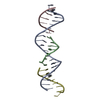
- PDB-5eos: Crystallizing Strained DNA: Self-Assembly of 3D Crystals Containi... -
+ Open data

- Basic information
| Entry | Database: PDB / ID: 5eos | ||||||
|---|---|---|---|---|---|---|---|
| Title | Crystallizing Strained DNA: Self-Assembly of 3D Crystals Containing a Torsionally-Stressed Component | ||||||
 Components Components |
| ||||||
 Keywords Keywords |  DNA / DNA nanotechnology / torsionally strained DNA / DNA lattices DNA / DNA nanotechnology / torsionally strained DNA / DNA lattices | ||||||
| Function / homology | DNA / DNA (> 10) Function and homology information Function and homology information | ||||||
| Biological species | synthetic construct (others) | ||||||
| Method | X-RAY DIFFRACTION / SYNCHROTRON / SAD / Resolution: 4.7 Å | ||||||
 Authors Authors | Hernandez, C. / Birktoft, J.J. / Seeman, N.C. | ||||||
 Citation Citation | Journal: Cell Chem Biol / Year: 2017 Title: Self-Assembly of 3D DNA Crystals Containing a Torsionally Stressed Component. Authors: Hernandez, C. / Birktoft, J.J. / Ohayon, Y.P. / Chandrasekaran, A.R. / Abdallah, H. / Sha, R. / Stojanoff, V. / Mao, C. / Seeman, N.C. | ||||||
| History |
|
- Structure visualization
| Structure viewer | Molecule: MolmilJmol/JSmol |
|---|
- Downloads & links
-Download
| PDBx/mmCIF format | 5eos.cif.gz | 29.3 KB | Display | PDBx/mmCIF format |
|---|---|---|---|---|
| PDB format | pdb5eos.ent.gz | 20.1 KB | Display | PDB format |
| PDBx/mmJSON format | 5eos.json.gz | Tree view | PDBx/mmJSON format | |
| Others |  Other downloads Other downloads |
-Validation report
| Arichive directory | https://data.pdbj.org/pub/pdb/validation_reports/eo/5eosftp://data.pdbj.org/pub/pdb/validation_reports/eo/5eos | HTTPS FTP |
|---|
-Related structure data
| Related structure data | |
|---|---|
| Similar structure data |
-Links
 PDBj
PDBj


- Assembly
| Deposited unit | 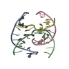
| ||||||||
|---|---|---|---|---|---|---|---|---|---|
| 1 | 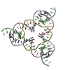
| ||||||||
| Unit cell |
| ||||||||
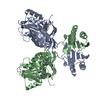
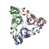
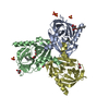
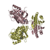
| Details | The asymmetric unit is comprised of the first 11 nucleotides of the full #1 strand (chain A), by the last 10 residues of the full #1 strand (chain C), by the first 8 nucleotides of the #2 strand (chain D) and by #3 strand (chain B). Chains A (First 11 residues of strand #1) and C (next 10 residues of strand #1) together form the first DNA strand (#1) of the experiment. This DNA strand spans two asymmetric units-hence it was divided into the current chains A and C for convenient representation and crystallographic computing. Chain A (1_555) is covalently linked to chain C of another asymmetric unit (3_555); and chain C (1_555) is covalently linked to chain A of another asymmetric unit (2_555). The #2 strand form a circular DNA molecule as a consequence of internal 3-fold symmetry of the triangle chain D in the asymmetric unit represents one of these repeating units. It has 3 symmetric and sequential repeating units that are covalently linked to each other via the O3' end of residue 13D to the O5' end of residue 6D; chain D (1_555) is covalently linked to chain D of two neighboring asymmetric units (2_555) and (3_555). The selection of residues from the DNA #2 and #3 are dictated by their proximity (base pairing) to chains A and C. Chain B in the asymmetric unit represents the third of these repeating units. |
-Components
| #1: DNA chain | Mass: 3414.234 Da / Num. of mol.: 1 Fragment: The first 11 residues the DNA molecule used in the experiment Source method: obtained synthetically / Source: (synth.) synthetic construct (others) |
|---|---|
| #2: DNA chain | Mass: 2371.582 Da / Num. of mol.: 1 / Source method: obtained synthetically / Source: (synth.) synthetic construct (others) |
| #3: DNA chain | Mass: 3973.582 Da / Num. of mol.: 1 / Source method: obtained synthetically / Source: (synth.) synthetic construct (others) |
| #4: DNA chain | Mass: 3038.020 Da / Num. of mol.: 1 Fragment: The second 10 residues the DNA molecule used in the experiment Source method: obtained synthetically / Source: (synth.) synthetic construct (others) |
-Experimental details
-Experiment
| Experiment | Method: X-RAY DIFFRACTION / Number of used crystals: 1 |
|---|
- Sample preparation
| Crystal | Density Matthews: 2.34 Å3/Da / Density % sol: 64.6 % / Description: rhombohedral |
|---|---|
| Crystal grow | Temperature: 277 K / Method: vapor diffusion, hanging drop / pH: 7 Details: Grown by vapor diffusion while treated with a controlled temperature gradient from 333 degs to 277 degs. Ammonium Sulfate, magnesium chloride, Na Cacodylate, magnesium acetate, TRIS PH range: 7 |
-Data collection
| Diffraction | Mean temperature: 100 K | |||||||||
|---|---|---|---|---|---|---|---|---|---|---|
| Diffraction source | Source: SYNCHROTRON / Site: NSLS  / Beamline: X25 / Wavelength: 1.1, 1.7 / Beamline: X25 / Wavelength: 1.1, 1.7 | |||||||||
| Detector | Type: PSI PILATUS 6M / Detector: PIXEL / Date: Aug 8, 2012 | |||||||||
| Radiation | Protocol: SINGLE WAVELENGTH / Monochromatic (M) / Laue (L): M / Scattering type: x-ray | |||||||||
| Radiation wavelength |
| |||||||||
| Reflection | Resolution: 4.7→27.5 Å / Num. obs: 4338 / % possible obs: 95 % / Redundancy: 1.8 % / Net I/σ(I): 2.6 | |||||||||
| Reflection shell | Resolution: 4.75→4.85 Å / Redundancy: 14.1 % / Rmerge(I) obs: 0.01 / Mean I/σ(I) obs: 2 / % possible all: 90 |
- Processing
| Software |
| ||||||||||||||||||
|---|---|---|---|---|---|---|---|---|---|---|---|---|---|---|---|---|---|---|---|
| Refinement | Method to determine structure: SAD / Resolution: 4.7→27.5 Å / SU ML: 0.87 / Cross valid method: FREE R-VALUE / σ(F): 2.03 / Phase error: 46.89
| ||||||||||||||||||
| Solvent computation | Shrinkage radii: 0.9 Å / VDW probe radii: 1.11 Å | ||||||||||||||||||
| Displacement parameters | Biso max: 371.51 Å2 / Biso mean: 317.8413 Å2 / Biso min: 272.89 Å2 | ||||||||||||||||||
| Refinement step | Cycle: final / Resolution: 4.7→27.5 Å
|
