 Movie
Movie Controller

[English] 日本語
 Yorodumi
- PDB-5emx: Crystal structure of the S. cerevisiae Rtf1 histone modification ... -
+ Open data

- Basic information
| Entry | Database: PDB / ID: 5emx | ||||||
|---|---|---|---|---|---|---|---|
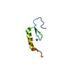
| Title | Crystal structure of the S. cerevisiae Rtf1 histone modification domain mutant R124A R126A R128A | ||||||
 Components Components | RNA polymerase-associated protein RTF1 | ||||||
 Keywords Keywords |  TRANSCRIPTION / Paf1 / Chromatin / Rtf1 TRANSCRIPTION / Paf1 / Chromatin / Rtf1 | ||||||
| Function / homology |  Function and homology information Function and homology informationsnoRNA transcription by RNA polymerase II / positive regulation of transcription elongation by RNA polymerase I / regulation of transcription-coupled nucleotide-excision repair / sno(s)RNA 3'-end processing / meiotic DNA double-strand break formation / RNA polymerase II C-terminal domain phosphoserine binding / Cdc73/Paf1 complex / global genome nucleotide-excision repair / mRNA 3'-end processing / : ...snoRNA transcription by RNA polymerase II / positive regulation of transcription elongation by RNA polymerase I / regulation of transcription-coupled nucleotide-excision repair / sno(s)RNA 3'-end processing / meiotic DNA double-strand break formation / RNA polymerase II C-terminal domain phosphoserine binding / Cdc73/Paf1 complex / global genome nucleotide-excision repair / mRNA 3'-end processing / : / transcription elongation by RNA polymerase II / DNA-templated transcription termination / positive regulation of transcription elongation by RNA polymerase II / euchromatin / regulation of transcription by RNA polymerase II / negative regulation of transcription by RNA polymerase II / DNA binding / RNA binding / nucleusSimilarity search - Function | ||||||
| Biological species |  Saccharomyces cerevisiae (brewer's yeast) Saccharomyces cerevisiae (brewer's yeast) | ||||||
| Method | X-RAY DIFFRACTION / SYNCHROTRON / SAD / Resolution: 1.399 Å | ||||||
 Authors Authors | Wier, A.D. / Heroux, A. / VanDemark, A.P. | ||||||
 Citation Citation | Journal: Mol.Cell / Year: 2016 Title: The Histone Modification Domain of Paf1 Complex Subunit Rtf1 Directly Stimulates H2B Ubiquitylation through an Interaction with Rad6. Authors: Van Oss, S.B. / Shirra, M.K. / Bataille, A.R. / Wier, A.D. / Yen, K. / Vinayachandran, V. / Byeon, I.L. / Cucinotta, C.E. / Heroux, A. / Jeon, J. / Kim, J. / VanDemark, A.P. / Pugh, B.F. / Arndt, K.M. | ||||||
| History |
|
- Structure visualization
| Structure viewer | Molecule: MolmilJmol/JSmol |
|---|
- Downloads & links
-Download
| PDBx/mmCIF format | 5emx.cif.gz | 80.5 KB | Display | PDBx/mmCIF format |
|---|---|---|---|---|
| PDB format | pdb5emx.ent.gz | 62.9 KB | Display | PDB format |
| PDBx/mmJSON format | 5emx.json.gz | Tree view | PDBx/mmJSON format | |
| Others |  Other downloads Other downloads |
-Validation report
| Arichive directory | https://data.pdbj.org/pub/pdb/validation_reports/em/5emxftp://data.pdbj.org/pub/pdb/validation_reports/em/5emx | HTTPS FTP |
|---|
-Related structure data


-Links
 PDBj
PDBj
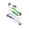
- Assembly
| Deposited unit | 
| ||||||||
|---|---|---|---|---|---|---|---|---|---|
| 1 | 
| ||||||||
| 2 | 
| ||||||||
| Unit cell |
| ||||||||
| Components on special symmetry positions |
|
-Components
| #1: Protein | Mass: 8516.430 Da / Num. of mol.: 2 / Fragment: UNP residues 74-139 / Mutation: R124A, R126A, R128A Source method: isolated from a genetically manipulated source Source: (gene. exp.) Saccharomyces cerevisiae (brewer's yeast)Strain: ATCC 204508 / S288c / Gene: RTF1, CSL3, YGL244W, HRA458 / Production host:  Escherichia coli (E. coli) / References: UniProt: P53064 Escherichia coli (E. coli) / References: UniProt: P53064#2: Water | ChemComp-HOH / | Water Mass: 18.015 Da / Num. of mol.: 78 / Source method: isolated from a natural source / Formula: H2O Mass: 18.015 Da / Num. of mol.: 78 / Source method: isolated from a natural source / Formula: H2O |
|---|
-Experimental details
-Experiment
| Experiment | Method: X-RAY DIFFRACTION / Number of used crystals: 1 |
|---|
- Sample preparation
| Crystal | Density Matthews: 1.93 Å3/Da / Density % sol: 36.24 % |
|---|---|
| Crystal grow | Temperature: 293 K / Method: vapor diffusion, sitting drop / Details: 0.1M phosphate-citrate (pH 4.2) and 40% PEG 300 |
-Data collection
| Diffraction | Mean temperature: 110 K |
|---|---|
| Diffraction source | Source: SYNCHROTRON / Site: NSLS  / Beamline: X25 / Wavelength: 1.1 Å / Beamline: X25 / Wavelength: 1.1 Å |
| Detector | Type: DECTRIS PILATUS 6M / Detector: PIXEL / Date: Jul 13, 2011 |
| Radiation | Protocol: SINGLE WAVELENGTH / Monochromatic (M) / Laue (L): M / Scattering type: x-ray |
| Radiation wavelength | Wavelength: 1.1 Å / Relative weight: 1 |
| Reflection | Resolution: 1.32→50 Å / Num. obs: 30553 / % possible obs: 98.3 % / Redundancy: 13.7 % / Rmerge(I) obs: 0.051 / Net I/σ(I): 77.79 |
| Reflection shell | Resolution: 1.32→1.34 Å / Redundancy: 3.5 % / Rmerge(I) obs: 0.626 / Mean I/σ(I) obs: 1.3 / % possible all: 78.5 |
- Processing
| Software |
| ||||||||||||||||||||||||||||||||||||||||||||||||||||||||
|---|---|---|---|---|---|---|---|---|---|---|---|---|---|---|---|---|---|---|---|---|---|---|---|---|---|---|---|---|---|---|---|---|---|---|---|---|---|---|---|---|---|---|---|---|---|---|---|---|---|---|---|---|---|---|---|---|---|
| Refinement | Method to determine structure: SAD / Resolution: 1.399→14.626 Å / SU ML: 0.11 / Cross valid method: THROUGHOUT / σ(F): 1.39 / Phase error: 17.77 / Stereochemistry target values: ML
| ||||||||||||||||||||||||||||||||||||||||||||||||||||||||
| Solvent computation | Shrinkage radii: 0.9 Å / VDW probe radii: 1.11 Å / Solvent model: FLAT BULK SOLVENT MODEL | ||||||||||||||||||||||||||||||||||||||||||||||||||||||||
| Refinement step | Cycle: LAST / Resolution: 1.399→14.626 Å
| ||||||||||||||||||||||||||||||||||||||||||||||||||||||||
| Refine LS restraints |
| ||||||||||||||||||||||||||||||||||||||||||||||||||||||||
| LS refinement shell |
|
