 Movie
Movie Controller


+ Open data

- Basic information
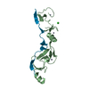
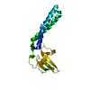
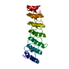
| Entry | Database: PDB / ID: 5eiq | ||||||
|---|---|---|---|---|---|---|---|
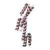
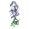
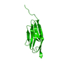
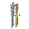
| Title | Human OSCAR ligand-binding domain | ||||||
 Components Components | Osteoclast-associated immunoglobulin-like receptor | ||||||
 Keywords Keywords |  IMMUNE SYSTEM / Immunoglobulin-like / collagen binding / immune receptor / osteoclast / BALBES NMR IMMUNE SYSTEM / Immunoglobulin-like / collagen binding / immune receptor / osteoclast / BALBES NMR | ||||||
| Function / homology |  Function and homology informationcollagen receptor activity / osteoclast differentiation / specific granule lumen / Immunoregulatory interactions between a Lymphoid and a non-Lymphoid cell / tertiary granule lumen / Neutrophil degranulation / extracellular exosome / extracellular region / plasma membrane Function and homology informationcollagen receptor activity / osteoclast differentiation / specific granule lumen / Immunoregulatory interactions between a Lymphoid and a non-Lymphoid cell / tertiary granule lumen / Neutrophil degranulation / extracellular exosome / extracellular region / plasma membraneSimilarity search - Function | ||||||
| Biological species |  Homo sapiens (human) Homo sapiens (human) | ||||||
| Method | X-RAY DIFFRACTION / SYNCHROTRON / MOLECULAR REPLACEMENT / Resolution: 2.01 Å | ||||||
 Authors Authors | Hinerman, J.M. / Conrady, D.G. / Herr, A.B. | ||||||
 Citation Citation | Journal: Blood / Year: 2016 Title: Structural basis for collagen recognition by the immune receptor OSCAR. Authors: Zhou, L. / Hinerman, J.M. / Blaszczyk, M. / Miller, J.L. / Conrady, D.G. / Barrow, A.D. / Chirgadze, D.Y. / Bihan, D. / Farndale, R.W. / Herr, A.B. | ||||||
| History |
|
- Structure visualization
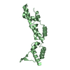
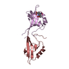
| Structure viewer | Molecule: MolmilJmol/JSmol |
|---|
- Downloads & links
-Download
| PDBx/mmCIF format | 5eiq.cif.gz | 91.2 KB | Display | PDBx/mmCIF format |
|---|---|---|---|---|
| PDB format | pdb5eiq.ent.gz | 67.6 KB | Display | PDB format |
| PDBx/mmJSON format | 5eiq.json.gz | Tree view | PDBx/mmJSON format | |
| Others |  Other downloads Other downloads |
-Validation report
| Arichive directory | https://data.pdbj.org/pub/pdb/validation_reports/ei/5eiqftp://data.pdbj.org/pub/pdb/validation_reports/ei/5eiq | HTTPS FTP |
|---|
-Related structure data
| Related structure data |  5eivC  3p2tS S: Starting model for refinement C: citing same article ( |
|---|---|
| Similar structure data |

-Links
 PDBj
PDBj
- Assembly
| Deposited unit | 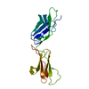
| ||||||||
|---|---|---|---|---|---|---|---|---|---|
| 1 |
| ||||||||
| Unit cell |
| ||||||||
| Details | Monomer confirmed by analytical ultracentrifugation |
-Components
| #1: Protein | Mass: 20731.713 Da / Num. of mol.: 1 Source method: isolated from a genetically manipulated source Source: (gene. exp.) Homo sapiens (human) / Gene: OSCAR / Plasmid: pDEST17 / Production host:  Escherichia coli (E. coli) / Strain (production host): BL21(DE3) / References: UniProt: Q8IYS5 Escherichia coli (E. coli) / Strain (production host): BL21(DE3) / References: UniProt: Q8IYS5 |
|---|---|
| #2: Water | ChemComp-HOH / Water Mass: 18.015 Da / Num. of mol.: 155 / Source method: isolated from a natural source / Formula: H2O Mass: 18.015 Da / Num. of mol.: 155 / Source method: isolated from a natural source / Formula: H2O |
-Experimental details
-Experiment
| Experiment | Method: X-RAY DIFFRACTION |
|---|
- Sample preparation
| Crystal | Density Matthews: 2.36 Å3/Da / Density % sol: 47.88 % |
|---|---|
| Crystal grow | Temperature: 277 K / Method: vapor diffusion / pH: 5 / Details: 0.1 M NaCitrate pH 5.0, 30% PEG 8000 |
-Data collection
| Diffraction | Mean temperature: 100 K | ||||||||||||||||||||||||||||||||||||||||||||||||||||||||||||||||||||||||||||||||||||||||||||||||||||||||||||||
|---|---|---|---|---|---|---|---|---|---|---|---|---|---|---|---|---|---|---|---|---|---|---|---|---|---|---|---|---|---|---|---|---|---|---|---|---|---|---|---|---|---|---|---|---|---|---|---|---|---|---|---|---|---|---|---|---|---|---|---|---|---|---|---|---|---|---|---|---|---|---|---|---|---|---|---|---|---|---|---|---|---|---|---|---|---|---|---|---|---|---|---|---|---|---|---|---|---|---|---|---|---|---|---|---|---|---|---|---|---|---|---|
| Diffraction source | Source: SYNCHROTRON / Site: APS  / Beamline: 24-ID-C / Wavelength: 0.97934 Å / Beamline: 24-ID-C / Wavelength: 0.97934 Å | ||||||||||||||||||||||||||||||||||||||||||||||||||||||||||||||||||||||||||||||||||||||||||||||||||||||||||||||
| Detector | Type: ADSC QUANTUM 315 / Detector: CCD / Date: Nov 11, 2011 | ||||||||||||||||||||||||||||||||||||||||||||||||||||||||||||||||||||||||||||||||||||||||||||||||||||||||||||||
| Radiation | Protocol: SINGLE WAVELENGTH / Monochromatic (M) / Laue (L): M / Scattering type: x-ray | ||||||||||||||||||||||||||||||||||||||||||||||||||||||||||||||||||||||||||||||||||||||||||||||||||||||||||||||
| Radiation wavelength | Wavelength: 0.97934 Å / Relative weight: 1 | ||||||||||||||||||||||||||||||||||||||||||||||||||||||||||||||||||||||||||||||||||||||||||||||||||||||||||||||
| Reflection | Resolution: 2.006→48.486 Å / Num. all: 12996 / Num. obs: 12996 / % possible obs: 99.1 % / Redundancy: 3.6 % / Biso Wilson estimate: 27.35 Å2 / Rpim(I) all: 0.1 / Rrim(I) all: 0.19 / Rsym value: 0.128 / Net I/av σ(I): 4.885 / Net I/σ(I): 7.7 / Num. measured all: 47065 | ||||||||||||||||||||||||||||||||||||||||||||||||||||||||||||||||||||||||||||||||||||||||||||||||||||||||||||||
| Reflection shell | Diffraction-ID: 1 / Rejects: 0
|
- Processing
| Software |
| |||||||||||||||||||||||||||||||||||||||||||||||||||||||||||||||||||||||||||||||||||||||||||||||||||||||||||||||||||||||||||||
|---|---|---|---|---|---|---|---|---|---|---|---|---|---|---|---|---|---|---|---|---|---|---|---|---|---|---|---|---|---|---|---|---|---|---|---|---|---|---|---|---|---|---|---|---|---|---|---|---|---|---|---|---|---|---|---|---|---|---|---|---|---|---|---|---|---|---|---|---|---|---|---|---|---|---|---|---|---|---|---|---|---|---|---|---|---|---|---|---|---|---|---|---|---|---|---|---|---|---|---|---|---|---|---|---|---|---|---|---|---|---|---|---|---|---|---|---|---|---|---|---|---|---|---|---|---|---|
| Refinement | Method to determine structure: MOLECULAR REPLACEMENT Starting model: 3P2T Resolution: 2.01→40.7 Å / Cor.coef. Fo:Fc: 0.9403 / Cor.coef. Fo:Fc free: 0.9113 / SU R Cruickshank DPI: 0.181 / Cross valid method: THROUGHOUT / σ(F): 0 / SU R Blow DPI: 0.191 / SU Rfree Blow DPI: 0.159 / SU Rfree Cruickshank DPI: 0.156
| |||||||||||||||||||||||||||||||||||||||||||||||||||||||||||||||||||||||||||||||||||||||||||||||||||||||||||||||||||||||||||||
| Displacement parameters | Biso max: 115.35 Å2 / Biso mean: 32.54 Å2 / Biso min: 7.84 Å2
| |||||||||||||||||||||||||||||||||||||||||||||||||||||||||||||||||||||||||||||||||||||||||||||||||||||||||||||||||||||||||||||
| Refine analyze | Luzzati coordinate error obs: 0.247 Å | |||||||||||||||||||||||||||||||||||||||||||||||||||||||||||||||||||||||||||||||||||||||||||||||||||||||||||||||||||||||||||||
| Refinement step | Cycle: final / Resolution: 2.01→40.7 Å
| |||||||||||||||||||||||||||||||||||||||||||||||||||||||||||||||||||||||||||||||||||||||||||||||||||||||||||||||||||||||||||||
| Refine LS restraints |
| |||||||||||||||||||||||||||||||||||||||||||||||||||||||||||||||||||||||||||||||||||||||||||||||||||||||||||||||||||||||||||||
| LS refinement shell | Resolution: 2.01→2.2 Å / Total num. of bins used: 6
| |||||||||||||||||||||||||||||||||||||||||||||||||||||||||||||||||||||||||||||||||||||||||||||||||||||||||||||||||||||||||||||
| Refinement TLS params. | Method: refined / Refine-ID: X-RAY DIFFRACTION
| |||||||||||||||||||||||||||||||||||||||||||||||||||||||||||||||||||||||||||||||||||||||||||||||||||||||||||||||||||||||||||||
| Refinement TLS group |
|
