 Movie
Movie Controller

[English] 日本語
 Yorodumi
- PDB-5eg7: The cap binding site of influenza virus protein PB2 as a drug target -
+ Open data

- Basic information
| Entry | Database: PDB / ID: 5eg7 | ||||||
|---|---|---|---|---|---|---|---|
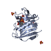
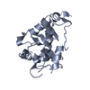
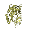
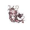
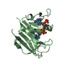
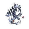
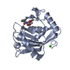
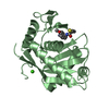
| Title | The cap binding site of influenza virus protein PB2 as a drug target | ||||||
 Components Components | Polymerase basic protein 2 | ||||||
 Keywords Keywords |  VIRAL PROTEIN / PB2 cap VIRAL PROTEIN / PB2 cap | ||||||
| Function / homology |  Function and homology informationcap snatching / symbiont-mediated suppression of host mRNA transcription via inhibition of RNA polymerase II activity / host cell mitochondrion / 7-methylguanosine mRNA capping / viral RNA genome replication / DNA-templated transcription / host cell nucleus / RNA binding Function and homology informationcap snatching / symbiont-mediated suppression of host mRNA transcription via inhibition of RNA polymerase II activity / host cell mitochondrion / 7-methylguanosine mRNA capping / viral RNA genome replication / DNA-templated transcription / host cell nucleus / RNA bindingSimilarity search - Function | ||||||
| Biological species |   Influenza A virus Influenza A virus | ||||||
| Method | X-RAY DIFFRACTION / SYNCHROTRON / MOLECULAR REPLACEMENT / Resolution: 1.4 Å | ||||||
 Authors Authors | Severin, C. / Rocha de Moura, T. / Liu, Y. / Li, K. / Zheng, X. / Luo, M. | ||||||
 Citation Citation | Journal: Acta Crystallogr D Struct Biol / Year: 2016 Title: The cap-binding site of influenza virus protein PB2 as a drug target. Authors: Severin, C. / Rocha de Moura, T. / Liu, Y. / Li, K. / Zheng, X. / Luo, M. | ||||||
| History |
|
- Structure visualization
| Structure viewer | Molecule: MolmilJmol/JSmol |
|---|
- Downloads & links
-Download
| PDBx/mmCIF format | 5eg7.cif.gz | 87.4 KB | Display | PDBx/mmCIF format |
|---|---|---|---|---|
| PDB format | pdb5eg7.ent.gz | 64.6 KB | Display | PDB format |
| PDBx/mmJSON format | 5eg7.json.gz | Tree view | PDBx/mmJSON format | |
| Others |  Other downloads Other downloads |
-Validation report
| Arichive directory | https://data.pdbj.org/pub/pdb/validation_reports/eg/5eg7ftp://data.pdbj.org/pub/pdb/validation_reports/eg/5eg7 | HTTPS FTP |
|---|
-Related structure data
| Related structure data |  5eg8SC  5eg9C S: Starting model for refinement C: citing same article ( |
|---|---|
| Similar structure data |


-Links
 PDBj
PDBj- Assembly
| Deposited unit | 
| ||||||||
|---|---|---|---|---|---|---|---|---|---|
| 1 |
| ||||||||
| Unit cell |
|
-Components
| #1: Protein | Mass: 18037.945 Da / Num. of mol.: 1 / Fragment: UNP residues 106-207, 215-269 Source method: isolated from a genetically manipulated source Source: (gene. exp.) Influenza A virus / Strain: A/swine/Quebec/1257777/2010(H3N2) / Gene: PB2 / Plasmid: pet28a / Production host:  Escherichia coli (E. coli) / Strain (production host): BL21 / References: UniProt: G3LZH2 Escherichia coli (E. coli) / Strain (production host): BL21 / References: UniProt: G3LZH2 |
|---|---|
| #2: Chemical | ChemComp-MGT /   Mass: 539.223 Da / Num. of mol.: 1 / Source method: obtained synthetically / Formula: C11H20N5O14P3 Mass: 539.223 Da / Num. of mol.: 1 / Source method: obtained synthetically / Formula: C11H20N5O14P3 |
| #3: Chemical | ChemComp-GOL / Glycerol  Mass: 92.094 Da / Num. of mol.: 1 / Source method: obtained synthetically / Formula: C3H8O3 Mass: 92.094 Da / Num. of mol.: 1 / Source method: obtained synthetically / Formula: C3H8O3 |
| #4: Water | ChemComp-HOH / Water Mass: 18.015 Da / Num. of mol.: 162 / Source method: isolated from a natural source / Formula: H2O Mass: 18.015 Da / Num. of mol.: 162 / Source method: isolated from a natural source / Formula: H2O |
-Experimental details
-Experiment
| Experiment | Method: X-RAY DIFFRACTION / Number of used crystals: 1 |
|---|
- Sample preparation
| Crystal | Density Matthews: 2.23 Å3/Da / Density % sol: 44.77 % |
|---|---|
| Crystal grow | Temperature: 293 K / Method: vapor diffusion / pH: 6.5 / Details: 10% PEG 3350 |
-Data collection
| Diffraction | Mean temperature: 100 K |
|---|---|
| Diffraction source | Source: SYNCHROTRON / Site: APS  / Beamline: 22-ID / Wavelength: 1 Å / Beamline: 22-ID / Wavelength: 1 Å |
| Detector | Type: MARMOSAIC 300 mm CCD / Detector: CCD / Date: Oct 26, 2015 |
| Radiation | Protocol: SINGLE WAVELENGTH / Monochromatic (M) / Laue (L): M / Scattering type: x-ray |
| Radiation wavelength | Wavelength: 1 Å / Relative weight: 1 |
| Reflection | Resolution: 1.4→31.356 Å / Num. obs: 31468 / % possible obs: 99.8 % / Redundancy: 10.4 % / Rsym value: 0.071 / Net I/σ(I): 2.3 |
- Processing
| Software |
| |||||||||||||||||||||||||||||||||||||||||||||||||||||||||||||||||||||||||||
|---|---|---|---|---|---|---|---|---|---|---|---|---|---|---|---|---|---|---|---|---|---|---|---|---|---|---|---|---|---|---|---|---|---|---|---|---|---|---|---|---|---|---|---|---|---|---|---|---|---|---|---|---|---|---|---|---|---|---|---|---|---|---|---|---|---|---|---|---|---|---|---|---|---|---|---|---|
| Refinement | Method to determine structure: MOLECULAR REPLACEMENT Starting model: 5EG8 Resolution: 1.4→31.356 Å / Cor.coef. Fo:Fc: 0.967 / Cor.coef. Fo:Fc free: 0.962 / SU B: 1.918 / SU ML: 0.037 / Cross valid method: THROUGHOUT / σ(F): 0 / ESU R: 0.059 / ESU R Free: 0.059 / Stereochemistry target values: MAXIMUM LIKELIHOOD Details: HYDROGENS HAVE BEEN ADDED IN THE RIDING POSITIONS U VALUES : WITH TLS ADDED
| |||||||||||||||||||||||||||||||||||||||||||||||||||||||||||||||||||||||||||
| Solvent computation | Ion probe radii: 0.8 Å / Shrinkage radii: 0.8 Å / VDW probe radii: 1.2 Å / Solvent model: MASK | |||||||||||||||||||||||||||||||||||||||||||||||||||||||||||||||||||||||||||
| Displacement parameters | Biso max: 77.12 Å2 / Biso mean: 14.531 Å2 / Biso min: 5.34 Å2
| |||||||||||||||||||||||||||||||||||||||||||||||||||||||||||||||||||||||||||
| Refinement step | Cycle: final / Resolution: 1.4→31.356 Å
| |||||||||||||||||||||||||||||||||||||||||||||||||||||||||||||||||||||||||||
| Refine LS restraints |
| |||||||||||||||||||||||||||||||||||||||||||||||||||||||||||||||||||||||||||
| LS refinement shell | Resolution: 1.4→1.436 Å / Total num. of bins used: 20
| |||||||||||||||||||||||||||||||||||||||||||||||||||||||||||||||||||||||||||
| Refinement TLS params. | Method: refined / Origin x: -21.1358 Å / Origin y: 5.8704 Å / Origin z: 11.4587 Å
|
