 Movie
Movie Controller

[English] 日本語
 Yorodumi
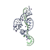
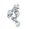
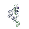
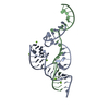
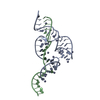
- PDB-5eaq: Two active site divalent ion in the crystal structure of the hamm... -
+ Open data

- Basic information
| Entry | Database: PDB / ID: 5eaq | ||||||
|---|---|---|---|---|---|---|---|
| Title | Two active site divalent ion in the crystal structure of the hammerhead ribozyme bound to a transition state analog-Mn2+ | ||||||
 Components Components |
| ||||||
 Keywords Keywords |  RNA / ribozyme / hammerhead RNA / ribozyme / hammerhead | ||||||
| Function / homology | : / RNA / RNA (> 10) Function and homology information Function and homology information | ||||||
| Biological species | synthetic construct (others) | ||||||
| Method | X-RAY DIFFRACTION / SYNCHROTRON / MOLECULAR REPLACEMENT / Resolution: 3.201 Å | ||||||
 Authors Authors | Mir, A. / Golden, B.L. | ||||||
 Citation Citation | Journal: Biochemistry / Year: 2016 Title: Two Active Site Divalent Ions in the Crystal Structure of the Hammerhead Ribozyme Bound to a Transition State Analogue. Authors: Mir, A. / Golden, B.L. #1: Journal: Biochemistry / Year: 2015Title: Two Divalent Metal Ions and Conformational Changes Play Roles in the Hammerhead Ribozyme Cleavage Reaction. Authors: Mir, A. / Chen, J. / Robinson, K. / Lendy, E. / Goodman, J. / Neau, D. / Golden, B.L. | ||||||
| History |
|
- Structure visualization
| Structure viewer | Molecule: MolmilJmol/JSmol |
|---|
- Downloads & links
-Download
| PDBx/mmCIF format | 5eaq.cif.gz | 48.8 KB | Display | PDBx/mmCIF format |
|---|---|---|---|---|
| PDB format | pdb5eaq.ent.gz | 34.4 KB | Display | PDB format |
| PDBx/mmJSON format | 5eaq.json.gz | Tree view | PDBx/mmJSON format | |
| Others |  Other downloads Other downloads |
-Validation report
| Arichive directory | https://data.pdbj.org/pub/pdb/validation_reports/ea/5eaqftp://data.pdbj.org/pub/pdb/validation_reports/ea/5eaq | HTTPS FTP |
|---|
-Related structure data
| Related structure data | 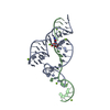 5eaoC 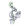 5di2S C: citing same article ( S: Starting model for refinement |
|---|---|
| Similar structure data |


-Links
 PDBj
PDBj


- Assembly
| Deposited unit | 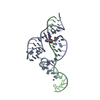
| ||||||||
|---|---|---|---|---|---|---|---|---|---|
| 1 |
| ||||||||
| Unit cell |
| ||||||||
| Components on special symmetry positions |
|
-Components
| #1: RNA chain | Mass: 15500.287 Da / Num. of mol.: 1 Source method: isolated from a genetically manipulated source Source: (gene. exp.) synthetic construct (others) / Plasmid: pUC19 / Production host:  Escherichia coli (E. coli) / Strain (production host): DH5alpha Escherichia coli (E. coli) / Strain (production host): DH5alpha |
|---|---|
| #2: RNA chain | Mass: 6536.834 Da / Num. of mol.: 1 / Source method: obtained synthetically / Source: (synth.) synthetic construct (others) |
| #3: Chemical | ChemComp-MN /   Mass: 54.938 Da / Num. of mol.: 6 / Source method: obtained synthetically / Formula: Mn Mass: 54.938 Da / Num. of mol.: 6 / Source method: obtained synthetically / Formula: Mn |
-Experimental details
-Experiment
| Experiment | Method: X-RAY DIFFRACTION |
|---|
- Sample preparation
| Crystal | Density Matthews: 4.25 Å3/Da / Density % sol: 71.07 % |
|---|---|
| Crystal grow | Temperature: 273 K / Method: vapor diffusion, hanging drop / pH: 8.2 Details: 50 mM Tris pH 8.2, 10 mM MgCl2, 32% MPD, 0.9 M KCl, 0.5 mM spermine 2 mM NH4VO3 |
-Data collection
| Diffraction | Mean temperature: 100 K |
|---|---|
| Diffraction source | Source: SYNCHROTRON / Site: APS  / Beamline: 21-ID-D / Wavelength: 1.501 Å / Beamline: 21-ID-D / Wavelength: 1.501 Å |
| Detector | Type: MARMOSAIC 300 mm CCD / Detector: CCD / Date: Nov 17, 2014 |
| Radiation | Protocol: SINGLE WAVELENGTH / Monochromatic (M) / Laue (L): M / Scattering type: x-ray |
| Radiation wavelength | Wavelength: 1.501 Å / Relative weight: 1 |
| Reflection | Resolution: 2.95→50 Å / Num. obs: 12773 / % possible obs: 99.4 % / Redundancy: 6.3 % / Rmerge(I) obs: 0.196 / Net I/σ(I): 139.7 |
| Reflection shell | Resolution: 2.95→3 Å / Redundancy: 5 % / Rmerge(I) obs: 0.636 / Mean I/σ(I) obs: 19.4 / % possible all: 97.2 |
- Processing
| Software |
| |||||||||||||||||||||||||||||||||||||||||||||||||||||||||||||||
|---|---|---|---|---|---|---|---|---|---|---|---|---|---|---|---|---|---|---|---|---|---|---|---|---|---|---|---|---|---|---|---|---|---|---|---|---|---|---|---|---|---|---|---|---|---|---|---|---|---|---|---|---|---|---|---|---|---|---|---|---|---|---|---|---|
| Refinement | Method to determine structure: MOLECULAR REPLACEMENT Starting model: 5DI2 Resolution: 3.201→27.429 Å / SU ML: 0.34 / Cross valid method: FREE R-VALUE / σ(F): 1.37 / Phase error: 30.95 / Stereochemistry target values: ML
| |||||||||||||||||||||||||||||||||||||||||||||||||||||||||||||||
| Solvent computation | Shrinkage radii: 0.9 Å / VDW probe radii: 1.11 Å / Solvent model: FLAT BULK SOLVENT MODEL | |||||||||||||||||||||||||||||||||||||||||||||||||||||||||||||||
| Refinement step | Cycle: LAST / Resolution: 3.201→27.429 Å
| |||||||||||||||||||||||||||||||||||||||||||||||||||||||||||||||
| Refine LS restraints |
| |||||||||||||||||||||||||||||||||||||||||||||||||||||||||||||||
| LS refinement shell |
|
