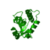
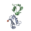
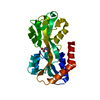
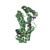
[English] 日本語
Yorodumi
- PDB-5e8g: Crystal structure of the DNA binding domain of human transcriptio... -

+
Open data


ID or keywords:

Loading...

-
Basic information

Entry
Database: PDB / ID: 5e8g
TitleCrystal structure of the DNA binding domain of human transcription factor FLI1
ComponentsFriend leukemia integration 1 transcription factor
KeywordsDNA BINDING PROTEIN / Transcription / DNA binding / Ewing sarcoma / Winged helix / ETS family
Function / homology
Function and homology information


hemostasis / blood circulation / megakaryocyte development / animal organ morphogenesis / Transcriptional regulation of granulopoiesis / sequence-specific double-stranded DNA binding / DNA-binding transcription activator activity, RNA polymerase II-specific / nuclear body / transcription cis-regulatory region binding / DNA-binding transcription factor activity, RNA polymerase II-specific ...hemostasis / blood circulation / megakaryocyte development / animal organ morphogenesis / Transcriptional regulation of granulopoiesis / sequence-specific double-stranded DNA binding / DNA-binding transcription activator activity, RNA polymerase II-specific / nuclear body / transcription cis-regulatory region binding / DNA-binding transcription factor activity, RNA polymerase II-specific / RNA polymerase II cis-regulatory region sequence-specific DNA binding / DNA-binding transcription factor activity / chromatin binding / chromatin / regulation of transcription by RNA polymerase II / positive regulation of DNA-templated transcription / DNA binding / nucleoplasm / nucleus / cytosol
Similarity search - Function
Friend leukemia integration 1 transcription factor, pointed domain / SAM / Pointed domain / Pointed domain / Sterile alpha motif (SAM)/Pointed domain / Pointed (PNT) domain profile. / Ets-domain signature 1. / ETS family / Ets-domain signature 2. / Ets domain / Ets-domain ...Friend leukemia integration 1 transcription factor, pointed domain / SAM / Pointed domain / Pointed domain / Sterile alpha motif (SAM)/Pointed domain / Pointed (PNT) domain profile. / Ets-domain signature 1. / ETS family / Ets-domain signature 2. / Ets domain / Ets-domain / Ets-domain profile. / erythroblast transformation specific domain / Sterile alpha motif/pointed domain superfamily / Winged helix-like DNA-binding domain superfamily/Winged helix DNA-binding domain / Arc Repressor Mutant, subunit A / Winged helix DNA-binding domain superfamily / Winged helix-like DNA-binding domain superfamily / Orthogonal Bundle / Mainly Alpha
Similarity search - Domain/homology
: / Friend leukemia integration 1 transcription factor
Similarity search - Component
Biological speciesHomo sapiens (human)
MethodX-RAY DIFFRACTION / SYNCHROTRON / MOLECULAR REPLACEMENT / Resolution: 2.7 Å
AuthorsHou, C. / Tsodikov, O.V.
CitationJournal: Biochemistry / Year: 2015
Title: Structural Basis for Dimerization and DNA Binding of Transcription Factor FLI1.
Authors: Hou, C. / Tsodikov, O.V.
History
DepositionOct 14, 2015Deposition site: RCSB / Processing site: RCSB
Revision 1.0Dec 9, 2015Provider: repository / Type: Initial release
Revision 1.1Dec 16, 2015Group: Database references
Revision 1.2Dec 30, 2015Group: Database references
Revision 1.3Nov 20, 2019Group: Database references / Derived calculations
Category: citation / pdbx_struct_conn_angle ...citation / pdbx_struct_conn_angle / pdbx_struct_oper_list / struct_conn
Item: _citation.journal_id_CSD / _pdbx_struct_oper_list.symmetry_operation
Revision 1.4Sep 27, 2023Group: Data collection / Database references ...Data collection / Database references / Derived calculations / Refinement description
Category: chem_comp_atom / chem_comp_bond ...chem_comp_atom / chem_comp_bond / database_2 / pdbx_initial_refinement_model / pdbx_struct_conn_angle / struct_conn
Item: _database_2.pdbx_DOI / _database_2.pdbx_database_accession ..._database_2.pdbx_DOI / _database_2.pdbx_database_accession / _pdbx_struct_conn_angle.ptnr1_auth_asym_id / _pdbx_struct_conn_angle.ptnr1_auth_comp_id / _pdbx_struct_conn_angle.ptnr1_auth_seq_id / _pdbx_struct_conn_angle.ptnr1_label_asym_id / _pdbx_struct_conn_angle.ptnr1_label_atom_id / _pdbx_struct_conn_angle.ptnr1_label_comp_id / _pdbx_struct_conn_angle.ptnr1_label_seq_id / _pdbx_struct_conn_angle.ptnr1_symmetry / _pdbx_struct_conn_angle.ptnr2_auth_asym_id / _pdbx_struct_conn_angle.ptnr2_label_asym_id / _pdbx_struct_conn_angle.ptnr3_auth_asym_id / _pdbx_struct_conn_angle.ptnr3_auth_comp_id / _pdbx_struct_conn_angle.ptnr3_auth_seq_id / _pdbx_struct_conn_angle.ptnr3_label_asym_id / _pdbx_struct_conn_angle.ptnr3_label_atom_id / _pdbx_struct_conn_angle.ptnr3_label_comp_id / _pdbx_struct_conn_angle.ptnr3_label_seq_id / _pdbx_struct_conn_angle.ptnr3_symmetry / _pdbx_struct_conn_angle.value / _struct_conn.pdbx_dist_value / _struct_conn.ptnr1_auth_asym_id / _struct_conn.ptnr1_auth_comp_id / _struct_conn.ptnr1_auth_seq_id / _struct_conn.ptnr1_label_asym_id / _struct_conn.ptnr1_label_atom_id / _struct_conn.ptnr1_label_comp_id / _struct_conn.ptnr1_label_seq_id / _struct_conn.ptnr1_symmetry / _struct_conn.ptnr2_auth_asym_id / _struct_conn.ptnr2_auth_comp_id / _struct_conn.ptnr2_auth_seq_id / _struct_conn.ptnr2_label_asym_id / _struct_conn.ptnr2_label_atom_id / _struct_conn.ptnr2_label_comp_id / _struct_conn.ptnr2_symmetry

-
Structure visualization

Structure viewerMolecule:
MolmilJmol/JSmol

Downloads & links

-
Assembly

Deposited unit
A: Friend leukemia integration 1 transcription factor
B: Friend leukemia integration 1 transcription factor
C: Friend leukemia integration 1 transcription factor
D: Friend leukemia integration 1 transcription factor
hetero molecules


Theoretical massNumber of molelcules
Total (without water)60,0238
Polymers59,7874
Non-polymers2364
Water21612
1
A: Friend leukemia integration 1 transcription factor
C: Friend leukemia integration 1 transcription factor
hetero molecules


Theoretical massNumber of molelcules
Total (without water)30,0124
Polymers29,8942
Non-polymers1182
Water362
TypeNameSymmetry operationNumber
identity operation1_555x,y,z1
Buried area1890 Å2
ΔGint-33 kcal/mol
Surface area10720 Å2
MethodPISA
2
B: Friend leukemia integration 1 transcription factor
D: Friend leukemia integration 1 transcription factor
hetero molecules


Theoretical massNumber of molelcules
Total (without water)30,0124
Polymers29,8942
Non-polymers1182
Water362
TypeNameSymmetry operationNumber
identity operation1_555x,y,z1
Buried area1890 Å2
ΔGint-32 kcal/mol
Surface area10590 Å2
MethodPISA
Unit cell
Length a, b, c (Å)140.551, 140.551, 85.140
Angle α, β, γ (deg.)90.00, 90.00, 120.00
Int Tables number146
Space group name H-MH3

-
Components

#1: Protein
Friend leukemia integration 1 transcription factor / Proto-oncogene Fli-1 / Transcription factor ERGB


Mass: 14946.829 Da / Num. of mol.: 4 / Fragment: UNP residues 276-399
Source method: isolated from a genetically manipulated source
Source: (gene. exp.) Homo sapiens (human) / Gene: FLI1 / Production host: Escherichia coli (E. coli) / Strain (production host): BL21(DE3) / References: UniProt: Q01543
#2: Chemical
ChemComp-CO / COBALT (II) ION


Mass: 58.933 Da / Num. of mol.: 4 / Source method: obtained synthetically / Formula: Co
#3: Water ChemComp-HOH / water / Water


Mass: 18.015 Da / Num. of mol.: 12 / Source method: isolated from a natural source / Formula: H2O

-
Experimental details

-
Experiment

ExperimentMethod: X-RAY DIFFRACTION / Number of used crystals: 1

-
Sample preparation

CrystalDensity Matthews: 2.71 Å3/Da / Density % sol: 54.56 %
Crystal growTemperature: 294 K / Method: vapor diffusion, hanging drop / pH: 5.5
Details: 0.1 M sodium acetate pH 5.5, 0.1 M Co2+ sulfate heptahydrate, 24% PEG 4000

-
Data collection

DiffractionMean temperature: 100 K
Diffraction sourceSource: SYNCHROTRON / Site: APS / Beamline: 21-ID-G / Wavelength: 0.978 Å
DetectorType: MARMOSAIC 300 mm CCD / Detector: CCD / Date: May 29, 2014
RadiationProtocol: SINGLE WAVELENGTH / Monochromatic (M) / Laue (L): M / Scattering type: x-ray
Radiation wavelengthWavelength: 0.978 Å / Relative weight: 1
ReflectionResolution: 2.7→50 Å / Num. obs: 17449 / % possible obs: 100 % / Redundancy: 4.6 % / Rmerge(I) obs: 0.09 / Net I/σ(I): 27
Reflection shellResolution: 2.7→2.75 Å / Redundancy: 4.7 % / Rmerge(I) obs: 0.67 / Mean I/σ(I) obs: 2.3 / % possible all: 100

-
Processing

Software
NameVersionClassification
REFMAC5.7.0032refinement
MOLREPphasing
HKL-2000data scaling
HKL-2000data reduction
RefinementMethod to determine structure: MOLECULAR REPLACEMENT
Starting model: 4IRG
Resolution: 2.7→40 Å / Cor.coef. Fo:Fc: 0.948 / Cor.coef. Fo:Fc free: 0.91 / SU B: 10.739 / SU ML: 0.223 / Cross valid method: THROUGHOUT / ESU R: 0.479 / ESU R Free: 0.297 / Stereochemistry target values: MAXIMUM LIKELIHOOD / Details: HYDROGENS HAVE BEEN ADDED IN THE RIDING POSITIONS
RfactorNum. reflection% reflectionSelection details
Rfree0.24835 869 5 %RANDOM
Rwork0.19637 ---
obs0.19896 16370 99.95 %-
Solvent computationIon probe radii: 0.8 Å / Shrinkage radii: 0.8 Å / VDW probe radii: 1.2 Å / Solvent model: MASK
Displacement parametersBiso mean: 69.26 Å2
Baniso -1Baniso -2Baniso -3
1--0.13 Å2-0.13 Å2-0 Å2
2---0.13 Å20 Å2
3---0.42 Å2
Refinement stepCycle: 1 / Resolution: 2.7→40 Å
ProteinNucleic acidLigandSolventTotal
Num. atoms3088 0 4 12 3104
Refine LS restraints
Refine-IDTypeDev idealDev ideal targetNumber
X-RAY DIFFRACTIONr_bond_refined_d0.0090.0193176
X-RAY DIFFRACTIONr_bond_other_d0.0020.022932
X-RAY DIFFRACTIONr_angle_refined_deg1.2331.9354264
X-RAY DIFFRACTIONr_angle_other_deg0.74836728
X-RAY DIFFRACTIONr_dihedral_angle_1_deg5.2365368
X-RAY DIFFRACTIONr_dihedral_angle_2_deg36.53623.571168
X-RAY DIFFRACTIONr_dihedral_angle_3_deg17.78715564
X-RAY DIFFRACTIONr_dihedral_angle_4_deg18.3191524
X-RAY DIFFRACTIONr_chiral_restr0.0710.2420
X-RAY DIFFRACTIONr_gen_planes_refined0.0040.023596
X-RAY DIFFRACTIONr_gen_planes_other0.0010.02812
X-RAY DIFFRACTIONr_nbd_refined
X-RAY DIFFRACTIONr_nbd_other
X-RAY DIFFRACTIONr_nbtor_refined
X-RAY DIFFRACTIONr_nbtor_other
X-RAY DIFFRACTIONr_xyhbond_nbd_refined
X-RAY DIFFRACTIONr_xyhbond_nbd_other
X-RAY DIFFRACTIONr_metal_ion_refined
X-RAY DIFFRACTIONr_metal_ion_other
X-RAY DIFFRACTIONr_symmetry_vdw_refined
X-RAY DIFFRACTIONr_symmetry_vdw_other
X-RAY DIFFRACTIONr_symmetry_hbond_refined
X-RAY DIFFRACTIONr_symmetry_hbond_other
X-RAY DIFFRACTIONr_symmetry_metal_ion_refined
X-RAY DIFFRACTIONr_symmetry_metal_ion_other
X-RAY DIFFRACTIONr_mcbond_it3.7346.6781484
X-RAY DIFFRACTIONr_mcbond_other3.7136.6761483
X-RAY DIFFRACTIONr_mcangle_it5.8589.9981848
X-RAY DIFFRACTIONr_mcangle_other5.857101849
X-RAY DIFFRACTIONr_scbond_it3.8147.1421692
X-RAY DIFFRACTIONr_scbond_other3.8137.1431693
X-RAY DIFFRACTIONr_scangle_it
X-RAY DIFFRACTIONr_scangle_other6.34410.542417
X-RAY DIFFRACTIONr_long_range_B_refined8.99553.6483731
X-RAY DIFFRACTIONr_long_range_B_other8.99453.6593732
X-RAY DIFFRACTIONr_rigid_bond_restr
X-RAY DIFFRACTIONr_sphericity_free
X-RAY DIFFRACTIONr_sphericity_bonded
LS refinement shellResolution: 2.7→2.77 Å / Total num. of bins used: 20
RfactorNum. reflection% reflection
Rfree0.366 68 -
Rwork0.272 1201 -
obs--100 %

+
About Yorodumi

-
News

-
Feb 9, 2022. New format data for meta-information of EMDB entries

New format data for meta-information of EMDB entries

  • Version 3 of the EMDB header file is now the official format.
  • The previous official version 1.9 will be removed from the archive.

Related info.:EMDB header

External links:wwPDB to switch to version 3 of the EMDB data model

-
Aug 12, 2020. Covid-19 info

Covid-19 info

URL: https://pdbj.org/emnavi/covid19.php

New page: Covid-19 featured information page in EM Navigator.

Related info.:Covid-19 info / Mar 5, 2020. Novel coronavirus structure data

+
Mar 5, 2020. Novel coronavirus structure data

Novel coronavirus structure data

Related info.:Yorodumi Speices / Aug 12, 2020. Covid-19 info

External links:COVID-19 featured content - PDBj / Molecule of the Month (242):Coronavirus Proteases

+
Jan 31, 2019. EMDB accession codes are about to change! (news from PDBe EMDB page)

EMDB accession codes are about to change! (news from PDBe EMDB page)

  • The allocation of 4 digits for EMDB accession codes will soon come to an end. Whilst these codes will remain in use, new EMDB accession codes will include an additional digit and will expand incrementally as the available range of codes is exhausted. The current 4-digit format prefixed with “EMD-” (i.e. EMD-XXXX) will advance to a 5-digit format (i.e. EMD-XXXXX), and so on. It is currently estimated that the 4-digit codes will be depleted around Spring 2019, at which point the 5-digit format will come into force.
  • The EM Navigator/Yorodumi systems omit the EMD- prefix.

Related info.:Q: What is EMD? / ID/Accession-code notation in Yorodumi/EM Navigator

External links:EMDB Accession Codes are Changing Soon! / Contact to PDBj

+
Jul 12, 2017. Major update of PDB

Major update of PDB

  • wwPDB released updated PDB data conforming to the new PDBx/mmCIF dictionary.
  • This is a major update changing the version number from 4 to 5, and with Remediation, in which all the entries are updated.
  • In this update, many items about electron microscopy experimental information are reorganized (e.g. em_software).
  • Now, EM Navigator and Yorodumi are based on the updated data.

External links:wwPDB Remediation / Enriched Model Files Conforming to OneDep Data Standards Now Available in the PDB FTP Archive

-
Yorodumi

Thousand views of thousand structures

  • Yorodumi is a browser for structure data from EMDB, PDB, SASBDB, etc.
  • This page is also the successor to EM Navigator detail page, and also detail information page/front-end page for Omokage search.
  • The word "yorodu" (or yorozu) is an old Japanese word meaning "ten thousand". "mi" (miru) is to see.

Related info.:EMDB / PDB / SASBDB / Comparison of 3 databanks / Yorodumi Search / Aug 31, 2016. New EM Navigator & Yorodumi / Yorodumi Papers / Jmol/JSmol / Function and homology information / Changes in new EM Navigator and Yorodumi

Read more