 Movie
Movie Controller

[English] 日本語
 Yorodumi
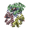
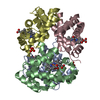
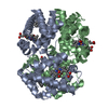
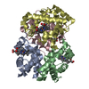
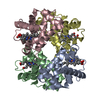
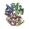
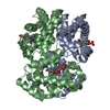
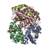
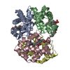
- PDB-5e83: CRYSTAL STRUCTURE OF CARBONMONOXY HEMOGLOBIN S (LIGANDED SICKLE C... -
+ Open data

- Basic information
| Entry | Database: PDB / ID: 5.0E+83 | ||||||
|---|---|---|---|---|---|---|---|
| Title | CRYSTAL STRUCTURE OF CARBONMONOXY HEMOGLOBIN S (LIGANDED SICKLE CELL HEMOGLOBIN) COMPLEXED WITH GBT440, CO-CRYSTALLIZATION EXPERIMENT | ||||||
 Components Components | (Hemoglobin subunit ... ) x 2 ) x 2 | ||||||
 Keywords Keywords | OXYGEN TRANSPORT / MUTANT HUMAN HEMOGLOBIN S[BETAE6V] / R2 QUATERNARY STATE | ||||||
| Function / homology |  Function and homology information Function and homology informationcellular oxidant detoxification / nitric oxide transport / hemoglobin alpha binding / haptoglobin-hemoglobin complex / hemoglobin binding / organic acid binding / renal absorption / hemoglobin complex / oxygen transport / Scavenging of heme from plasma ...cellular oxidant detoxification / nitric oxide transport / hemoglobin alpha binding / haptoglobin-hemoglobin complex / hemoglobin binding / organic acid binding / renal absorption / hemoglobin complex / oxygen transport / Scavenging of heme from plasma / endocytic vesicle lumen / blood vessel diameter maintenance / hydrogen peroxide catabolic process / oxygen carrier activity / Late endosomal microautophagy / Heme signaling / carbon dioxide transport / Erythrocytes take up oxygen and release carbon dioxide / response to hydrogen peroxide / Erythrocytes take up carbon dioxide and release oxygen / Cytoprotection by HMOX1 / platelet aggregation / oxygen binding / regulation of blood pressure / Chaperone Mediated Autophagy / positive regulation of nitric oxide biosynthetic process / tertiary granule lumen / Factors involved in megakaryocyte development and platelet production / ficolin-1-rich granule lumen / blood microparticle / iron ion binding / heme binding / Neutrophil degranulation / extracellular space / extracellular exosome / extracellular region / membrane / metal ion binding / cytosolSimilarity search - Function | ||||||
| Biological species |  Homo sapiens (human) Homo sapiens (human) | ||||||
| Method | X-RAY DIFFRACTION / SYNCHROTRON / MOLECULAR REPLACEMENT / Resolution: 1.8 Å | ||||||
 Authors Authors | Patskovska, L. / Patskovsky, Y. / Bonanno, J.B. / Almo, S.C. | ||||||
 Citation Citation | Journal: Br.J.Haematol. / Year: 2016 Title: GBT440 increases haemoglobin oxygen affinity, reduces sickling and prolongs RBC half-life in a murine model of sickle cell disease. Authors: Oksenberg, D. / Dufu, K. / Patel, M.P. / Chuang, C. / Li, Z. / Xu, Q. / Silva-Garcia, A. / Zhou, C. / Hutchaleelaha, A. / Patskovska, L. / Patskovsky, Y. / Almo, S.C. / Sinha, U. / Metcalf, B.W. / Archer, D.R. | ||||||
| History |
|
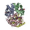
- Structure visualization
| Structure viewer | Molecule: MolmilJmol/JSmol |
|---|
- Downloads & links
-Download
| PDBx/mmCIF format | 5e83.cif.gz | 255.6 KB | Display | PDBx/mmCIF format |
|---|---|---|---|---|
| PDB format | pdb5e83.ent.gz | 205.8 KB | Display | PDB format |
| PDBx/mmJSON format | 5e83.json.gz | Tree view | PDBx/mmJSON format | |
| Others |  Other downloads Other downloads |
-Validation report
| Arichive directory | https://data.pdbj.org/pub/pdb/validation_reports/e8/5e83ftp://data.pdbj.org/pub/pdb/validation_reports/e8/5e83 | HTTPS FTP |
|---|
-Related structure data
| Related structure data | 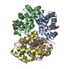 1nejS S: Starting model for refinement |
|---|---|
| Similar structure data |

-Links
 PDBj
PDBj


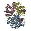
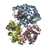
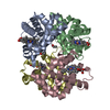
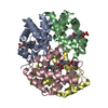
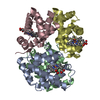
- Assembly
| Deposited unit | 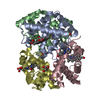
| ||||||||||||||||||||||||||||||||||||||||||||||||||||||||||||
|---|---|---|---|---|---|---|---|---|---|---|---|---|---|---|---|---|---|---|---|---|---|---|---|---|---|---|---|---|---|---|---|---|---|---|---|---|---|---|---|---|---|---|---|---|---|---|---|---|---|---|---|---|---|---|---|---|---|---|---|---|---|
| 1 |
| ||||||||||||||||||||||||||||||||||||||||||||||||||||||||||||
| Unit cell |
| ||||||||||||||||||||||||||||||||||||||||||||||||||||||||||||
| Noncrystallographic symmetry (NCS) | NCS domain:
NCS domain segments:
NCS ensembles :
NCS oper:
|
-Components
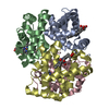
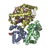
-Hemoglobin subunit ... , 2 types, 4 molecules ACBD
| #1: Protein | / Alpha-globin / Hemoglobin alpha chain Mass: 15150.353 Da / Num. of mol.: 2 / Source method: isolated from a natural source / Source: (natural) Homo sapiens (human) / References: UniProt: P69905#2: Protein | / Beta-globin / Hemoglobin beta chainMass: 15860.216 Da / Num. of mol.: 2 / Source method: isolated from a natural source / Source: (natural) Homo sapiens (human) / References: UniProt: P68871 |
|---|
-Non-polymers , 6 types, 412 molecules 


| #3: Chemical | ChemComp-HEM / Heme B Mass: 616.487 Da / Num. of mol.: 4 / Source method: obtained synthetically / Formula: C34H32FeN4O4 Mass: 616.487 Da / Num. of mol.: 4 / Source method: obtained synthetically / Formula: C34H32FeN4O4#4: Chemical | ChemComp-CMO / Carbon monoxide Mass: 28.010 Da / Num. of mol.: 4 / Source method: obtained synthetically / Formula: CO Mass: 28.010 Da / Num. of mol.: 4 / Source method: obtained synthetically / Formula: CO#5: Chemical | ChemComp-5L7 / |  Mass: 323.389 Da / Num. of mol.: 1 / Source method: obtained synthetically / Formula: C19H21N3O2 Mass: 323.389 Da / Num. of mol.: 1 / Source method: obtained synthetically / Formula: C19H21N3O2#6: Chemical | Glycerol Mass: 92.094 Da / Num. of mol.: 3 / Source method: obtained synthetically / Formula: C3H8O3 Mass: 92.094 Da / Num. of mol.: 3 / Source method: obtained synthetically / Formula: C3H8O3#7: Chemical | ChemComp-UNL / | Mass: 103.120 Da / Num. of mol.: 1 / Source method: obtained synthetically #8: Water | ChemComp-HOH / | WaterMass: 18.015 Da / Num. of mol.: 399 / Source method: isolated from a natural source / Formula: H2O |
|---|
-Experimental details
-Experiment
| Experiment | Method: X-RAY DIFFRACTION / Number of used crystals: 1 |
|---|
- Sample preparation
| Crystal | Density Matthews: 2.17 Å3/Da / Density % sol: 48.41 % |
|---|---|
| Crystal grow | Temperature: 294 K / Method: vapor diffusion, sitting drop / pH: 7.4 / Details: 0.1M HEPES, 25% PEG4000, PH 7.4, 40 MM NACL |
-Data collection
| Diffraction | Mean temperature: 100 K |
|---|---|
| Diffraction source | Source: SYNCHROTRON / Site: NSLS  / Beamline: X29A / Wavelength: 1.075 Å / Beamline: X29A / Wavelength: 1.075 Å |
| Detector | Type: ADSC QUANTUM 315 / Detector: CCD / Date: Jul 18, 2013 / Details: MIRRORS |
| Radiation | Monochromator: ROSENBAUM-ROCK DOUBLE CRYSTAL / Protocol: SINGLE WAVELENGTH / Monochromatic (M) / Laue (L): M / Scattering type: x-ray |
| Radiation wavelength | Wavelength: 1.075 Å / Relative weight: 1 |
| Reflection | Resolution: 1.8→50 Å / Num. obs: 56423 / % possible obs: 99.1 % / Observed criterion σ(I): -3 / Redundancy: 7.7 % / Rsym value: 0.063 / Net I/σ(I): 12.4 |
| Reflection shell | Resolution: 1.8→1.82 Å / Redundancy: 7.2 % / Rmerge(I) obs: 0.57 / Mean I/σ(I) obs: 3 / Rejects: 0 / % possible all: 98.6 |
- Processing
| Software |
| |||||||||||||||||||||||||||||||||||||||||||||||||||||||||||||||||||||||||||||||||||||||||||||||||||||||||||||||||||||||||||||
|---|---|---|---|---|---|---|---|---|---|---|---|---|---|---|---|---|---|---|---|---|---|---|---|---|---|---|---|---|---|---|---|---|---|---|---|---|---|---|---|---|---|---|---|---|---|---|---|---|---|---|---|---|---|---|---|---|---|---|---|---|---|---|---|---|---|---|---|---|---|---|---|---|---|---|---|---|---|---|---|---|---|---|---|---|---|---|---|---|---|---|---|---|---|---|---|---|---|---|---|---|---|---|---|---|---|---|---|---|---|---|---|---|---|---|---|---|---|---|---|---|---|---|---|---|---|---|
| Refinement | Method to determine structure: MOLECULAR REPLACEMENT Starting model: PDB ENTRY 1NEJ Resolution: 1.8→41.71 Å / Cor.coef. Fo:Fc: 0.969 / Cor.coef. Fo:Fc free: 0.959 / SU B: 6.791 / SU ML: 0.103 / Cross valid method: THROUGHOUT / σ(F): 0 / ESU R: 0.125 / ESU R Free: 0.116 / Stereochemistry target values: MAXIMUM LIKELIHOOD Details: HYDROGENS HAVE BEEN ADDED IN THE RIDING POSITIONS U VALUES : WITH TLS ADDED
| |||||||||||||||||||||||||||||||||||||||||||||||||||||||||||||||||||||||||||||||||||||||||||||||||||||||||||||||||||||||||||||
| Solvent computation | Ion probe radii: 0.8 Å / Shrinkage radii: 0.8 Å / VDW probe radii: 1.3 Å / Solvent model: BABINET MODEL WITH MASK | |||||||||||||||||||||||||||||||||||||||||||||||||||||||||||||||||||||||||||||||||||||||||||||||||||||||||||||||||||||||||||||
| Displacement parameters | Biso max: 126.52 Å2 / Biso mean: 40.129 Å2 / Biso min: 18.28 Å2
| |||||||||||||||||||||||||||||||||||||||||||||||||||||||||||||||||||||||||||||||||||||||||||||||||||||||||||||||||||||||||||||
| Refinement step | Cycle: final / Resolution: 1.8→41.71 Å
| |||||||||||||||||||||||||||||||||||||||||||||||||||||||||||||||||||||||||||||||||||||||||||||||||||||||||||||||||||||||||||||
| Refine LS restraints |
| |||||||||||||||||||||||||||||||||||||||||||||||||||||||||||||||||||||||||||||||||||||||||||||||||||||||||||||||||||||||||||||
| Refine LS restraints NCS | Dom-ID: 1 / Refine-ID: X-RAY DIFFRACTION
| |||||||||||||||||||||||||||||||||||||||||||||||||||||||||||||||||||||||||||||||||||||||||||||||||||||||||||||||||||||||||||||
| LS refinement shell | Resolution: 1.8→1.847 Å / Total num. of bins used: 20
| |||||||||||||||||||||||||||||||||||||||||||||||||||||||||||||||||||||||||||||||||||||||||||||||||||||||||||||||||||||||||||||
| Refinement TLS params. | Method: refined / Refine-ID: X-RAY DIFFRACTION
| |||||||||||||||||||||||||||||||||||||||||||||||||||||||||||||||||||||||||||||||||||||||||||||||||||||||||||||||||||||||||||||
| Refinement TLS group |
|
