 Movie
Movie Controller

[English] 日本語
 Yorodumi
- PDB-5e2a: Crystal structure of NTMT1 in complex with N-terminally methylate... -
+ Open data

- Basic information
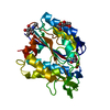
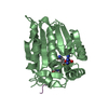
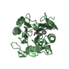
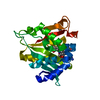
| Entry | Database: PDB / ID: 5e2a | ||||||
|---|---|---|---|---|---|---|---|
| Title | Crystal structure of NTMT1 in complex with N-terminally methylated SPKRIA peptide | ||||||
 Components Components |
| ||||||
 Keywords Keywords |  TRANSFERASE / methyl transferase / Structural Genomics / Structural Genomics Consortium / SGC TRANSFERASE / methyl transferase / Structural Genomics / Structural Genomics Consortium / SGC | ||||||
| Function / homology |  Function and homology information Function and homology informationN-terminal peptidyl-glycine methylation / N-terminal peptidyl-serine dimethylation / N-terminal peptidyl-serine trimethylation / protein N-terminal methyltransferase / N-terminal peptidyl-proline dimethylation / N-terminal protein N-methyltransferase activity / mitotic nuclear membrane reassembly / protein methyltransferase activity / sulfate binding / Rev-mediated nuclear export of HIV RNA ...N-terminal peptidyl-glycine methylation / N-terminal peptidyl-serine dimethylation / N-terminal peptidyl-serine trimethylation / protein N-terminal methyltransferase / N-terminal peptidyl-proline dimethylation / N-terminal protein N-methyltransferase activity / mitotic nuclear membrane reassembly / protein methyltransferase activity / sulfate binding / Rev-mediated nuclear export of HIV RNA / Nuclear import of Rev protein / Postmitotic nuclear pore complex (NPC) reformation / regulation of mitotic nuclear division / spindle organization / histone methyltransferase activity / nucleosomal DNA binding / nucleosome binding / spindle assembly / viral process / guanyl-nucleotide exchange factor activity / mitotic spindle organization / condensed nuclear chromosome / chromosome segregation / G1/S transition of mitotic cell cycle / small GTPase binding / chromosome / histone binding / protein heterodimerization activity / cell division / chromatin binding / chromatin / protein-containing complex / nucleoplasm / nucleus / cytosol / cytoplasmSimilarity search - Function | ||||||
| Biological species |  Homo sapiens (human) Homo sapiens (human) | ||||||
| Method | X-RAY DIFFRACTION / FOURIER SYNTHESIS / Resolution: 1.75 Å | ||||||
 Authors Authors | Dong, C. / Tempel, W. / Bountra, C. / Arrowsmith, C.H. / Edwards, A.M. / Min, J. / Structural Genomics Consortium (SGC) | ||||||
 Citation Citation | Journal: Genes Dev. / Year: 2015 Title: Structural basis for substrate recognition by the human N-terminal methyltransferase 1. Authors: Dong, C. / Mao, Y. / Tempel, W. / Qin, S. / Li, L. / Loppnau, P. / Huang, R. / Min, J. | ||||||
| History |
|
- Structure visualization
| Structure viewer | Molecule: MolmilJmol/JSmol |
|---|
- Downloads & links
-Download
| PDBx/mmCIF format | 5e2a.cif.gz | 209.3 KB | Display | PDBx/mmCIF format |
|---|---|---|---|---|
| PDB format | pdb5e2a.ent.gz | 165.4 KB | Display | PDB format |
| PDBx/mmJSON format | 5e2a.json.gz | Tree view | PDBx/mmJSON format | |
| Others |  Other downloads Other downloads |
-Validation report
| Arichive directory | https://data.pdbj.org/pub/pdb/validation_reports/e2/5e2aftp://data.pdbj.org/pub/pdb/validation_reports/e2/5e2a | HTTPS FTP |
|---|
-Related structure data
| Related structure data |  5e1bC  5e1dC  5e1mC  5e1oC  5e2bC  2ex4S C: citing same article ( S: Starting model for refinement |
|---|---|
| Similar structure data |


-Links
 PDBj
PDBj

- Assembly
| Deposited unit | 
| ||||||||
|---|---|---|---|---|---|---|---|---|---|
| 1 | 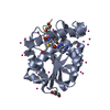
| ||||||||
| 2 | 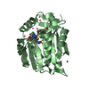
| ||||||||
| Unit cell |
|
-Components
-Protein / Protein/peptide , 2 types, 4 molecules ABDE
| #1: Protein | Mass: 27320.074 Da / Num. of mol.: 2 Source method: isolated from a genetically manipulated source Source: (gene. exp.) Homo sapiens (human) / Gene: NTMT1, C9orf32, METTL11A, NRMT, NRMT1, AD-003 / Plasmid: pET28a-LIC / Production host:  Escherichia coli (E. coli) / Strain (production host): BL21(DE3)-V3R Escherichia coli (E. coli) / Strain (production host): BL21(DE3)-V3RReferences: UniProt: Q9BV86, protein N-terminal methyltransferase#2: Protein/peptide | Mass: 686.843 Da / Num. of mol.: 2 / Source method: obtained synthetically / Details: synthetic peptide / Source: (synth.) Homo sapiens (human) / References: UniProt: P18754*PLUS |
|---|
-Non-polymers , 4 types, 465 molecules 


| #3: Chemical | S-Adenosyl-L-homocysteine Type: L-peptide linking / Mass: 384.411 Da / Num. of mol.: 2 / Source method: obtained synthetically / Formula: C14H20N6O5S Type: L-peptide linking / Mass: 384.411 Da / Num. of mol.: 2 / Source method: obtained synthetically / Formula: C14H20N6O5S#4: Chemical | ChemComp-GOL / Glycerol Mass: 92.094 Da / Num. of mol.: 4 / Source method: obtained synthetically / Formula: C3H8O3 Mass: 92.094 Da / Num. of mol.: 4 / Source method: obtained synthetically / Formula: C3H8O3#5: Chemical | ChemComp-UNX /  Num. of mol.: 33 / Source method: obtained synthetically Num. of mol.: 33 / Source method: obtained synthetically#6: Water | ChemComp-HOH / | WaterMass: 18.015 Da / Num. of mol.: 426 / Source method: isolated from a natural source / Formula: H2O |
|---|
-Experimental details
-Experiment
| Experiment | Method: X-RAY DIFFRACTION / Number of used crystals: 1 |
|---|
- Sample preparation
| Crystal | Density Matthews: 2.98 Å3/Da / Density % sol: 58.79 % |
|---|---|
| Crystal grow | Temperature: 277 K / Method: vapor diffusion, sitting drop / Details: 26% PEG3350, 16% tacsimate |
-Data collection
| Diffraction | Mean temperature: 100 K | ||||||||||||||||||||||||||||||
|---|---|---|---|---|---|---|---|---|---|---|---|---|---|---|---|---|---|---|---|---|---|---|---|---|---|---|---|---|---|---|---|
| Diffraction source | Source: ROTATING ANODE / Type: RIGAKU FR-E SUPERBRIGHT / Wavelength: 1.5418 Å | ||||||||||||||||||||||||||||||
| Detector | Type: RIGAKU SATURN A200 / Detector: CCD / Date: Aug 8, 2015 | ||||||||||||||||||||||||||||||
| Radiation | Protocol: SINGLE WAVELENGTH / Monochromatic (M) / Laue (L): M / Scattering type: x-ray | ||||||||||||||||||||||||||||||
| Radiation wavelength | Wavelength: 1.5418 Å / Relative weight: 1 | ||||||||||||||||||||||||||||||
| Reflection | Resolution: 1.75→29.62 Å / Num. obs: 70896 / % possible obs: 100 % / Redundancy: 20.8 % / CC1/2: 0.999 / Rmerge(I) obs: 0.125 / Rpim(I) all: 0.028 / Rrim(I) all: 0.129 / Net I/σ(I): 27.6 / Num. measured all: 1473712 | ||||||||||||||||||||||||||||||
| Reflection shell | Diffraction-ID: 1 / Rejects: 0
|
- Processing
| Software |
| |||||||||||||||||||||||||||||||||||||||||||||||||||||||||||||||||||||||||||
|---|---|---|---|---|---|---|---|---|---|---|---|---|---|---|---|---|---|---|---|---|---|---|---|---|---|---|---|---|---|---|---|---|---|---|---|---|---|---|---|---|---|---|---|---|---|---|---|---|---|---|---|---|---|---|---|---|---|---|---|---|---|---|---|---|---|---|---|---|---|---|---|---|---|---|---|---|
| Refinement | Method to determine structure: FOURIER SYNTHESIS Starting model: pdbid 2ex4 Resolution: 1.75→29.6 Å / Cor.coef. Fo:Fc: 0.961 / Cor.coef. Fo:Fc free: 0.944 / WRfactor Rfree: 0.161 / WRfactor Rwork: 0.138 / FOM work R set: 0.8996 / SU B: 2.969 / SU ML: 0.053 / SU R Cruickshank DPI: 0.0808 / SU Rfree: 0.0819 / Cross valid method: THROUGHOUT / σ(F): 0 / ESU R: 0.081 / ESU R Free: 0.082 / Stereochemistry target values: MAXIMUM LIKELIHOOD Details: Coot was used for interactive model building. Molprobity was used for geometry validation.
| |||||||||||||||||||||||||||||||||||||||||||||||||||||||||||||||||||||||||||
| Solvent computation | Ion probe radii: 0.8 Å / Shrinkage radii: 0.8 Å / VDW probe radii: 1.2 Å / Solvent model: MASK | |||||||||||||||||||||||||||||||||||||||||||||||||||||||||||||||||||||||||||
| Displacement parameters | Biso max: 48.69 Å2 / Biso mean: 17.264 Å2 / Biso min: 7.13 Å2
| |||||||||||||||||||||||||||||||||||||||||||||||||||||||||||||||||||||||||||
| Refinement step | Cycle: final / Resolution: 1.75→29.6 Å
| |||||||||||||||||||||||||||||||||||||||||||||||||||||||||||||||||||||||||||
| Refine LS restraints |
| |||||||||||||||||||||||||||||||||||||||||||||||||||||||||||||||||||||||||||
| LS refinement shell | Resolution: 1.75→1.795 Å / Total num. of bins used: 20
| |||||||||||||||||||||||||||||||||||||||||||||||||||||||||||||||||||||||||||
| Refinement TLS params. | Method: refined / Refine-ID: X-RAY DIFFRACTION
| |||||||||||||||||||||||||||||||||||||||||||||||||||||||||||||||||||||||||||
| Refinement TLS group |
|
