 Movie
Movie Controller

[English] 日本語
 Yorodumi
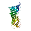
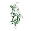
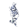
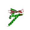
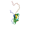
- PDB-5dyr: Structure of virulence-associated protein D (VapD) from Xylella f... -
+ Open data

- Basic information
| Entry | Database: PDB / ID: 5dyr | ||||||
|---|---|---|---|---|---|---|---|
| Title | Structure of virulence-associated protein D (VapD) from Xylella fastidiosa | ||||||
 Components Components | Virulence-associated protein D | ||||||
 Keywords Keywords |  DNA BINDING PROTEIN / VapD / Virulence-associated protein D / Xylella fastidiosa / cell invasion DNA BINDING PROTEIN / VapD / Virulence-associated protein D / Xylella fastidiosa / cell invasion | ||||||
| Function / homology | Virulence-associated protein D / CRISPR associated protein Cas2 / CRISPR associated protein Cas2 / nuclease activity / Virulence-associated protein D Function and homology information Function and homology information | ||||||
| Biological species |  Xylella fastidiosa (bacteria) Xylella fastidiosa (bacteria) | ||||||
| Method | X-RAY DIFFRACTION / SYNCHROTRON / MOLECULAR REPLACEMENT / Resolution: 3 Å | ||||||
 Authors Authors | Kochneva, M.V. / dos Santos, M.L. / dos Santos, C.A. / de Souza, A.P. / Polikarpov, I. / Aparicio, R. / Golubev, A.M. | ||||||
 Citation Citation | Journal: To Be Published Title: Structure of virulence-associated protein D (VapD) from Xylella fastidiosa Authors: Kochneva, M.V. / dos Santos, M.L. / dos Santos, C.A. / de Souza, A.P. / Polikarpov, I. / Aparicio, R. / Golubev, A.M. | ||||||
| History |
|
- Structure visualization
| Structure viewer | Molecule: MolmilJmol/JSmol |
|---|
- Downloads & links
-Download
| PDBx/mmCIF format | 5dyr.cif.gz | 41.6 KB | Display | PDBx/mmCIF format |
|---|---|---|---|---|
| PDB format | pdb5dyr.ent.gz | 29.3 KB | Display | PDB format |
| PDBx/mmJSON format | 5dyr.json.gz | Tree view | PDBx/mmJSON format | |
| Others |  Other downloads Other downloads |
-Validation report
| Arichive directory | https://data.pdbj.org/pub/pdb/validation_reports/dy/5dyrftp://data.pdbj.org/pub/pdb/validation_reports/dy/5dyr | HTTPS FTP |
|---|
-Related structure data
| Similar structure data |
|---|


-Links
 PDBj
PDBj- Assembly
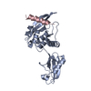
| Deposited unit | 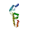
| ||||||||
|---|---|---|---|---|---|---|---|---|---|
| 1 |
| ||||||||
| Unit cell |
|
-Components
| #1: Protein | Mass: 17658.842 Da / Num. of mol.: 1 Source method: isolated from a genetically manipulated source Source: (gene. exp.) Xylella fastidiosa (strain 9a5c) (bacteria)Strain: 9a5c / Gene: XF_a0052 / Plasmid: pET32Xa/LIC / Production host: Escherichia coli BL21(DE3) (bacteria) / References: UniProt: Q9PHF6 |
|---|---|
| #2: Water | ChemComp-HOH / Water Mass: 18.015 Da / Num. of mol.: 8 / Source method: isolated from a natural source / Formula: H2O Mass: 18.015 Da / Num. of mol.: 8 / Source method: isolated from a natural source / Formula: H2O |
-Experimental details
-Experiment
| Experiment | Method: X-RAY DIFFRACTION |
|---|
- Sample preparation
| Crystal grow | Temperature: 289 K / Method: vapor diffusion, hanging drop / pH: 4.5 / Details: 20% MPD, 0.1M NaAc pH 4.5, 170mM glycine |
|---|
-Data collection
| Diffraction | Mean temperature: 100 K |
|---|---|
| Diffraction source | Source: SYNCHROTRON / Site: LNLS  / Beamline: W01B-MX2 / Wavelength: 1.4586 Å / Beamline: W01B-MX2 / Wavelength: 1.4586 Å |
| Detector | Type: MARMOSAIC 225 mm CCD / Detector: CCD / Date: Sep 11, 2009 |
| Radiation | Protocol: SINGLE WAVELENGTH / Monochromatic (M) / Laue (L): M / Scattering type: x-ray |
| Radiation wavelength | Wavelength: 1.4586 Å / Relative weight: 1 |
| Reflection | Resolution: 2.2→35.1 Å / Num. obs: 15925 / % possible obs: 85.8 % / Redundancy: 11.4 % / Rsym value: 0.218 / Net I/σ(I): 6.7 |
- Processing
| Software |
| ||||||||||||||||||||||||||||||||||||||||||||||||||||||||||||||||||||||||||||||||||||||||||||||||||||||||||||||||||||||||||||||||||||||||||||||||||||||||||||||||||||||||||||||||||||||
|---|---|---|---|---|---|---|---|---|---|---|---|---|---|---|---|---|---|---|---|---|---|---|---|---|---|---|---|---|---|---|---|---|---|---|---|---|---|---|---|---|---|---|---|---|---|---|---|---|---|---|---|---|---|---|---|---|---|---|---|---|---|---|---|---|---|---|---|---|---|---|---|---|---|---|---|---|---|---|---|---|---|---|---|---|---|---|---|---|---|---|---|---|---|---|---|---|---|---|---|---|---|---|---|---|---|---|---|---|---|---|---|---|---|---|---|---|---|---|---|---|---|---|---|---|---|---|---|---|---|---|---|---|---|---|---|---|---|---|---|---|---|---|---|---|---|---|---|---|---|---|---|---|---|---|---|---|---|---|---|---|---|---|---|---|---|---|---|---|---|---|---|---|---|---|---|---|---|---|---|---|---|---|---|
| Refinement | Method to determine structure: MOLECULAR REPLACEMENT / Resolution: 3→18.69 Å / Cor.coef. Fo:Fc: 0.927 / Cor.coef. Fo:Fc free: 0.897 / SU B: 7.291 / SU ML: 0.151 / Cross valid method: THROUGHOUT / ESU R: 0.123 / ESU R Free: 0.075 / Stereochemistry target values: MAXIMUM LIKELIHOOD / Details: HYDROGENS HAVE BEEN ADDED IN THE RIDING POSITIONS
| ||||||||||||||||||||||||||||||||||||||||||||||||||||||||||||||||||||||||||||||||||||||||||||||||||||||||||||||||||||||||||||||||||||||||||||||||||||||||||||||||||||||||||||||||||||||
| Solvent computation | Ion probe radii: 0.8 Å / Shrinkage radii: 0.8 Å / VDW probe radii: 1.2 Å / Solvent model: MASK | ||||||||||||||||||||||||||||||||||||||||||||||||||||||||||||||||||||||||||||||||||||||||||||||||||||||||||||||||||||||||||||||||||||||||||||||||||||||||||||||||||||||||||||||||||||||
| Displacement parameters | Biso mean: 60.259 Å2
| ||||||||||||||||||||||||||||||||||||||||||||||||||||||||||||||||||||||||||||||||||||||||||||||||||||||||||||||||||||||||||||||||||||||||||||||||||||||||||||||||||||||||||||||||||||||
| Refinement step | Cycle: LAST / Resolution: 3→18.69 Å
| ||||||||||||||||||||||||||||||||||||||||||||||||||||||||||||||||||||||||||||||||||||||||||||||||||||||||||||||||||||||||||||||||||||||||||||||||||||||||||||||||||||||||||||||||||||||
| Refine LS restraints |
|