 Movie
Movie Controller


+ Open data

- Basic information
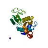
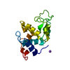
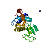
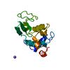
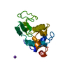
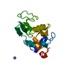
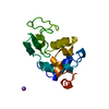
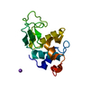
| Entry | Database: PDB / ID: 5dps | ||||||
|---|---|---|---|---|---|---|---|
| Title | Crystal structure of PLEKHM1 LIR-fused human GABARAP_2-117 | ||||||
 Components Components | Pleckstrin homology domain-containing family M member 1,Gamma-aminobutyric acid receptor-associated protein | ||||||
 Keywords Keywords |  PROTEIN BINDING / Autophagy / chimeric protein PROTEIN BINDING / Autophagy / chimeric protein | ||||||
| Function / homology |  Function and homology information Function and homology informationpositive regulation of protein K48-linked ubiquitination / late endosome to lysosome transport / autophagosome-lysosome fusion / regulation of Rac protein signal transduction / autolysosome membrane / GABA receptor binding / lysosome localization / cellular response to nitrogen starvation / phosphatidylethanolamine binding / positive regulation of ruffle assembly ...positive regulation of protein K48-linked ubiquitination / late endosome to lysosome transport / autophagosome-lysosome fusion / regulation of Rac protein signal transduction / autolysosome membrane / GABA receptor binding / lysosome localization / cellular response to nitrogen starvation / phosphatidylethanolamine binding / positive regulation of ruffle assembly / TBC/RABGAPs / microtubule associated complex / autolysosome / Macroautophagy / beta-tubulin binding / smooth endoplasmic reticulum / axoneme / autophagosome membrane / autophagosome assembly / extrinsic apoptotic signaling pathway via death domain receptors / protein targeting / autophagosome / positive regulation of bone resorption / sperm midpiece / macroautophagy / microtubule cytoskeleton organization / actin cytoskeleton / positive regulation of proteasomal ubiquitin-dependent protein catabolic process / protein transport / late endosome membrane / cytoplasmic vesicle / microtubule binding / chemical synaptic transmission / microtubule / lysosome / Golgi membrane / intracellular membrane-bounded organelle / synapse / ubiquitin protein ligase binding / nucleolus / metal ion binding / plasma membrane / cytosolSimilarity search - Function | ||||||
| Biological species |  Homo sapiens (human) Homo sapiens (human) | ||||||
| Method | X-RAY DIFFRACTION / SYNCHROTRON / MOLECULAR REPLACEMENT / Resolution: 2 Å | ||||||
 Authors Authors | Ravichandran, A.C. / Suzuki, H. / Dobson, R.C.J. | ||||||
 Citation Citation | Journal: EMBO Rep. / Year: 2017 Title: Structural and functional analysis of the GABARAP interaction motif (GIM). Authors: Rogov, V.V. / Stolz, A. / Ravichandran, A.C. / Rios-Szwed, D.O. / Suzuki, H. / Kniss, A. / Lohr, F. / Wakatsuki, S. / Dotsch, V. / Dikic, I. / Dobson, R.C. / McEwan, D.G. | ||||||
| History |
|
- Structure visualization
| Structure viewer | Molecule: MolmilJmol/JSmol |
|---|
- Downloads & links
-Download
| PDBx/mmCIF format | 5dps.cif.gz | 90 KB | Display | PDBx/mmCIF format |
|---|---|---|---|---|
| PDB format | pdb5dps.ent.gz | 67.5 KB | Display | PDB format |
| PDBx/mmJSON format | 5dps.json.gz | Tree view | PDBx/mmJSON format | |
| Others |  Other downloads Other downloads |
-Validation report
| Arichive directory | https://data.pdbj.org/pub/pdb/validation_reports/dp/5dpsftp://data.pdbj.org/pub/pdb/validation_reports/dp/5dps | HTTPS FTP |
|---|
-Related structure data


-Links
 PDBj
PDBj


- Assembly
| Deposited unit | 
| ||||||||
|---|---|---|---|---|---|---|---|---|---|
| 1 | 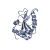
| ||||||||
| 2 | 
| ||||||||
| 3 | 
| ||||||||
| Unit cell |
|
-Components
| #1: Protein | Mass: 15580.702 Da / Num. of mol.: 3 Fragment: UNP Q9Y4G2 residues 627-638, UNP O95166 residues 2-117 Source method: isolated from a genetically manipulated source Source: (gene. exp.) Homo sapiens (human) / Gene: PLEKHM1, GABARAP, / Plasmid: pET30 / Cell line (production host): BL21(de3) / Production host:  Escherichia coli (E. coli) / References: UniProt: Q9Y4G2, UniProt: O95166 Escherichia coli (E. coli) / References: UniProt: Q9Y4G2, UniProt: O95166#2: Water | ChemComp-HOH / | Water Mass: 18.015 Da / Num. of mol.: 177 / Source method: isolated from a natural source / Formula: H2O Mass: 18.015 Da / Num. of mol.: 177 / Source method: isolated from a natural source / Formula: H2O |
|---|
-Experimental details
-Experiment
| Experiment | Method: X-RAY DIFFRACTION / Number of used crystals: 1 |
|---|
- Sample preparation
| Crystal | Density Matthews: 2.5 Å3/Da / Density % sol: 50.89 % |
|---|---|
| Crystal grow | Temperature: 278 K / Method: vapor diffusion, sitting drop / pH: 8 Details: 4% v/v Tacsimate pH 8.0, 12% w/v Polyethylene glycol 3,350 |
-Data collection
| Diffraction | Mean temperature: 100 K |
|---|---|
| Diffraction source | Source: SYNCHROTRON / Site: Australian Synchrotron  / Beamline: MX2 / Wavelength: 0.9537 Å / Beamline: MX2 / Wavelength: 0.9537 Å |
| Detector | Type: ADSC QUANTUM 315r / Detector: CCD / Date: Jul 26, 2014 |
| Radiation | Protocol: SINGLE WAVELENGTH / Monochromatic (M) / Laue (L): M / Scattering type: x-ray |
| Radiation wavelength | Wavelength: 0.9537 Å / Relative weight: 1 |
| Reflection | Resolution: 2→36.903 Å / Num. obs: 30464 / % possible obs: 99.2 % / Redundancy: 11.6 % / Net I/σ(I): 10.3 |
| Reflection shell | Resolution: 2→2.05 Å / % possible all: 98.6 |
- Processing
| Software |
| ||||||||||||||||||||||||||||||||||||||||||||||||||||||||||||||||||||||||||||||||||||
|---|---|---|---|---|---|---|---|---|---|---|---|---|---|---|---|---|---|---|---|---|---|---|---|---|---|---|---|---|---|---|---|---|---|---|---|---|---|---|---|---|---|---|---|---|---|---|---|---|---|---|---|---|---|---|---|---|---|---|---|---|---|---|---|---|---|---|---|---|---|---|---|---|---|---|---|---|---|---|---|---|---|---|---|---|---|
| Refinement | Method to determine structure: MOLECULAR REPLACEMENT / Resolution: 2→36.903 Å / SU ML: 0.26 / Cross valid method: FREE R-VALUE / σ(F): 1.98 / Phase error: 26.79 / Stereochemistry target values: ML
| ||||||||||||||||||||||||||||||||||||||||||||||||||||||||||||||||||||||||||||||||||||
| Solvent computation | Shrinkage radii: 0.9 Å / VDW probe radii: 1.11 Å / Solvent model: FLAT BULK SOLVENT MODEL | ||||||||||||||||||||||||||||||||||||||||||||||||||||||||||||||||||||||||||||||||||||
| Refinement step | Cycle: LAST / Resolution: 2→36.903 Å
| ||||||||||||||||||||||||||||||||||||||||||||||||||||||||||||||||||||||||||||||||||||
| Refine LS restraints |
| ||||||||||||||||||||||||||||||||||||||||||||||||||||||||||||||||||||||||||||||||||||
| LS refinement shell |
|
