[English] 日本語
Yorodumi
- PDB-5dob: Crystal structure of the Human Cytomegalovirus Nuclear Egress Com... -

+
Open data


ID or keywords:

Loading...

-
Basic information

Entry
Database: PDB / ID: 5dob
TitleCrystal structure of the Human Cytomegalovirus Nuclear Egress Complex (NEC)
Components
  • Virion egress protein UL31 homolog
  • Virion egress protein UL34 homolog
Keywords
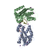
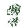
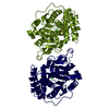
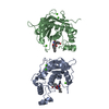
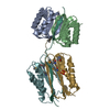
DNA BINDING PROTEIN / nuclear egress complex / zinc finger / interaction interface / Bergerat fold
Function / homology
Function and homology information


host cell nuclear inner membrane / viral budding from nuclear membrane / membrane / metal ion binding
Similarity search - Function
Herpesvirus viron egress-type / Herpesvirus virion protein U34 / Herpesvirus UL31 / Herpesvirus UL31-like protein
Similarity search - Domain/homology
Nuclear egress protein 2 / Nuclear egress protein 1
Similarity search - Component
Biological speciesHuman cytomegalovirus
MethodX-RAY DIFFRACTION / SYNCHROTRON / Resolution: 2.47 Å
AuthorsLye, M.F. / El Omari, K. / Filman, D.J. / Hogle, J.M.
Funding support United States, 1items
OrganizationGrant numberCountry
National Institutes of Health/National Institute Of Allergy and Infectious Diseases (NIH/NIAID)R01 AI026077 United States
CitationJournal: Embo J. / Year: 2015
Title: Unexpected features and mechanism of heterodimer formation of a herpesvirus nuclear egress complex.
Authors: Lye, M.F. / Sharma, M. / El Omari, K. / Filman, D.J. / Schuermann, J.P. / Hogle, J.M. / Coen, D.M.
History
DepositionSep 11, 2015Deposition site: RCSB / Processing site: RCSB
Revision 1.0Nov 25, 2015Provider: repository / Type: Initial release
Revision 1.1Dec 16, 2015Group: Database references
Revision 1.2Sep 20, 2017Group: Author supporting evidence / Database references ...Author supporting evidence / Database references / Derived calculations / Refinement description
Category: citation / pdbx_audit_support ...citation / pdbx_audit_support / pdbx_struct_oper_list / software
Item: _citation.journal_id_CSD / _pdbx_audit_support.funding_organization ..._citation.journal_id_CSD / _pdbx_audit_support.funding_organization / _pdbx_struct_oper_list.symmetry_operation / _software.classification
Revision 1.3Dec 11, 2019Group: Author supporting evidence / Category: pdbx_audit_support / Item: _pdbx_audit_support.funding_organization
Revision 1.4Mar 6, 2024Group: Data collection / Database references / Derived calculations
Category: chem_comp_atom / chem_comp_bond ...chem_comp_atom / chem_comp_bond / database_2 / struct_conn
Item: _database_2.pdbx_DOI / _database_2.pdbx_database_accession ..._database_2.pdbx_DOI / _database_2.pdbx_database_accession / _struct_conn.pdbx_dist_value / _struct_conn.ptnr1_auth_comp_id / _struct_conn.ptnr1_auth_seq_id / _struct_conn.ptnr1_label_atom_id / _struct_conn.ptnr1_label_comp_id / _struct_conn.ptnr1_label_seq_id / _struct_conn.ptnr2_auth_comp_id / _struct_conn.ptnr2_auth_seq_id / _struct_conn.ptnr2_label_asym_id / _struct_conn.ptnr2_label_atom_id / _struct_conn.ptnr2_label_comp_id / _struct_conn.ptnr2_symmetry

-
Structure visualization

Structure viewerMolecule:
MolmilJmol/JSmol

Downloads & links

-
Assembly

Deposited unit
A: Virion egress protein UL31 homolog
B: Virion egress protein UL34 homolog
hetero molecules


Theoretical massNumber of molelcules
Total (without water)45,7344
Polymers45,6292
Non-polymers1052
Water0
1


  • Idetical with deposited unit
  • defined by author&software
TypeNameSymmetry operationNumber
identity operation1_555x,y,z1
Buried area3980 Å2
ΔGint-36 kcal/mol
Surface area20260 Å2
MethodPISA
Unit cell
Length a, b, c (Å)142.254, 33.978, 92.995
Angle α, β, γ (deg.)90.00, 121.76, 90.00
Int Tables number5
Space group name H-MC121

-
Components

#1: Protein Virion egress protein UL31 homolog


Mass: 27176.248 Da / Num. of mol.: 1 / Fragment: unp residues 61-289 / Mutation: H62S, D63E, I67V, R69Q, and E70R
Source method: isolated from a genetically manipulated source
Source: (gene. exp.) Human cytomegalovirus / Strain: AD169 / Gene: UL53 / Production host: Escherichia coli (E. coli) / References: UniProt: P16794
#2: Protein Virion egress protein UL34 homolog / Primary envelopment factor UL34 homolog


Mass: 18452.348 Da / Num. of mol.: 1 / Fragment: unp residues 4-168
Source method: isolated from a genetically manipulated source
Source: (gene. exp.) Human cytomegalovirus (strain AD169) / Strain: AD169 / Gene: UL50 / Production host: Escherichia coli (E. coli) / References: UniProt: P16791
#3: Chemical ChemComp-ZN / ZINC ION


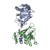
Mass: 65.409 Da / Num. of mol.: 1 / Source method: obtained synthetically / Formula: Zn
#4: Chemical ChemComp-CA / CALCIUM ION


Mass: 40.078 Da / Num. of mol.: 1 / Source method: obtained synthetically / Formula: Ca

-
Experimental details

-
Experiment

ExperimentMethod: X-RAY DIFFRACTION

-
Sample preparation

CrystalDensity Matthews: 2.09 Å3/Da / Density % sol: 41.26 %
Crystal growTemperature: 298 K / Method: vapor diffusion, hanging drop / pH: 7.9
Details: 0.1 M Tris, 0.1 M NaCl, 10-15% PEG 3350 (w/v), 0.3-0.5 M CaCl2

-
Data collection

DiffractionMean temperature: 100 K
Diffraction sourceSource: SYNCHROTRON / Site: APS / Beamline: 24-ID-E / Wavelength: 0.9792 Å
DetectorType: ADSC QUANTUM 315 / Detector: CCD / Date: Jun 10, 2012
RadiationProtocol: SINGLE WAVELENGTH / Monochromatic (M) / Laue (L): M / Scattering type: x-ray
Radiation wavelengthWavelength: 0.9792 Å / Relative weight: 1
ReflectionResolution: 2.47→79.08 Å / Num. obs: 13720 / % possible obs: 97.5 % / Redundancy: 3.5 % / Net I/σ(I): 14.47
Reflection shellHighest resolution: 2.47 Å / Redundancy: 2.1 % / % possible all: 88.9

-
Processing

Software
NameVersionClassification
REFMAC5.8.0049refinement
XDSdata scaling
PHASERphasing
RefinementResolution: 2.47→79.07 Å / Cor.coef. Fo:Fc: 0.931 / Cor.coef. Fo:Fc free: 0.891 / SU B: 42.84 / SU ML: 0.392 / Cross valid method: THROUGHOUT / ESU R: 3.174 / ESU R Free: 0.378 / Stereochemistry target values: MAXIMUM LIKELIHOOD / Details: HYDROGENS HAVE BEEN ADDED IN THE RIDING POSITIONS
RfactorNum. reflection% reflectionSelection details
Rfree0.2992 698 5.1 %RANDOM
Rwork0.25423 ---
obs0.25644 12896 96.91 %-
Solvent computationIon probe radii: 0.8 Å / Shrinkage radii: 0.8 Å / VDW probe radii: 1.2 Å / Solvent model: MASK
Displacement parametersBiso mean: 88.923 Å2
Baniso -1Baniso -2Baniso -3
1--3.61 Å20 Å22.97 Å2
2---8.47 Å20 Å2
3---4.76 Å2
Refinement stepCycle: LAST / Resolution: 2.47→79.07 Å
ProteinNucleic acidLigandSolventTotal
Num. atoms3189 0 2 0 3191
Refine LS restraints
Refine-IDTypeDev idealDev ideal targetNumber
X-RAY DIFFRACTIONr_bond_refined_d0.0050.0193254
X-RAY DIFFRACTIONr_bond_other_d0.0010.023121
X-RAY DIFFRACTIONr_angle_refined_deg1.0191.9614412
X-RAY DIFFRACTIONr_angle_other_deg0.70237190
X-RAY DIFFRACTIONr_dihedral_angle_1_deg5.0715400
X-RAY DIFFRACTIONr_dihedral_angle_2_deg33.22724.362149
X-RAY DIFFRACTIONr_dihedral_angle_3_deg12.69415574
X-RAY DIFFRACTIONr_dihedral_angle_4_deg13.7531518
X-RAY DIFFRACTIONr_chiral_restr0.0580.2509
X-RAY DIFFRACTIONr_gen_planes_refined0.0040.0213648
X-RAY DIFFRACTIONr_gen_planes_other0.0010.02734
X-RAY DIFFRACTIONr_nbd_refined
X-RAY DIFFRACTIONr_nbd_other
X-RAY DIFFRACTIONr_nbtor_refined
X-RAY DIFFRACTIONr_nbtor_other
X-RAY DIFFRACTIONr_xyhbond_nbd_refined
X-RAY DIFFRACTIONr_xyhbond_nbd_other
X-RAY DIFFRACTIONr_metal_ion_refined
X-RAY DIFFRACTIONr_metal_ion_other
X-RAY DIFFRACTIONr_symmetry_vdw_refined
X-RAY DIFFRACTIONr_symmetry_vdw_other
X-RAY DIFFRACTIONr_symmetry_hbond_refined
X-RAY DIFFRACTIONr_symmetry_hbond_other
X-RAY DIFFRACTIONr_symmetry_metal_ion_refined
X-RAY DIFFRACTIONr_symmetry_metal_ion_other
X-RAY DIFFRACTIONr_mcbond_it1.8845.9541606
X-RAY DIFFRACTIONr_mcbond_other1.8845.9541605
X-RAY DIFFRACTIONr_mcangle_it3.3568.9182004
X-RAY DIFFRACTIONr_mcangle_other3.3558.9192005
X-RAY DIFFRACTIONr_scbond_it1.4926.1591648
X-RAY DIFFRACTIONr_scbond_other1.4916.1591649
X-RAY DIFFRACTIONr_scangle_it
X-RAY DIFFRACTIONr_scangle_other2.7149.1492409
X-RAY DIFFRACTIONr_long_range_B_refined6.5546.2543440
X-RAY DIFFRACTIONr_long_range_B_other6.54946.2583441
X-RAY DIFFRACTIONr_rigid_bond_restr
X-RAY DIFFRACTIONr_sphericity_free
X-RAY DIFFRACTIONr_sphericity_bonded
LS refinement shellResolution: 2.469→2.533 Å / Total num. of bins used: 20
RfactorNum. reflection% reflection
Rfree0.387 45 -
Rwork0.38 762 -
obs--79.74 %
Refinement TLS params.

Method: refined / Refine-ID: X-RAY DIFFRACTION

IDL112)L122)L132)L222)L232)L332)S11 (Å °)S12 (Å °)S13 (Å °)S21 (Å °)S22 (Å °)S23 (Å °)S31 (Å °)S32 (Å °)S33 (Å °)T112)T122)T132)T222)T232)T332)Origin x (Å)Origin y (Å)Origin z (Å)
10.88220.10431.18110.60660.62432.13230.12940.01160.1432-0.2794-0.1493-0.23980.01120.06010.01990.3008-0.00030.17470.51770.09120.1428-26.7766-1.3766-38.194
24.82560.5824-0.35650.5956-0.02980.03350.0857-0.0559-0.01540.016-0.0899-0.0386-0.02860.04330.00420.143-0.0175-0.00940.48150.0370.0051-22.8508-3.7487-3.8331
Refinement TLS group
IDRefine-IDRefine TLS-IDAuth asym-IDAuth seq-ID
1X-RAY DIFFRACTION1A53 - 289
2X-RAY DIFFRACTION2B4 - 168

+
About Yorodumi

-
News

-
Feb 9, 2022. New format data for meta-information of EMDB entries

New format data for meta-information of EMDB entries

  • Version 3 of the EMDB header file is now the official format.
  • The previous official version 1.9 will be removed from the archive.

Related info.:EMDB header

External links:wwPDB to switch to version 3 of the EMDB data model

-
Aug 12, 2020. Covid-19 info

Covid-19 info

URL: https://pdbj.org/emnavi/covid19.php

New page: Covid-19 featured information page in EM Navigator.

Related info.:Covid-19 info / Mar 5, 2020. Novel coronavirus structure data

+
Mar 5, 2020. Novel coronavirus structure data

Novel coronavirus structure data

Related info.:Yorodumi Speices / Aug 12, 2020. Covid-19 info

External links:COVID-19 featured content - PDBj / Molecule of the Month (242):Coronavirus Proteases

+
Jan 31, 2019. EMDB accession codes are about to change! (news from PDBe EMDB page)

EMDB accession codes are about to change! (news from PDBe EMDB page)

  • The allocation of 4 digits for EMDB accession codes will soon come to an end. Whilst these codes will remain in use, new EMDB accession codes will include an additional digit and will expand incrementally as the available range of codes is exhausted. The current 4-digit format prefixed with “EMD-” (i.e. EMD-XXXX) will advance to a 5-digit format (i.e. EMD-XXXXX), and so on. It is currently estimated that the 4-digit codes will be depleted around Spring 2019, at which point the 5-digit format will come into force.
  • The EM Navigator/Yorodumi systems omit the EMD- prefix.

Related info.:Q: What is EMD? / ID/Accession-code notation in Yorodumi/EM Navigator

External links:EMDB Accession Codes are Changing Soon! / Contact to PDBj

+
Jul 12, 2017. Major update of PDB

Major update of PDB

  • wwPDB released updated PDB data conforming to the new PDBx/mmCIF dictionary.
  • This is a major update changing the version number from 4 to 5, and with Remediation, in which all the entries are updated.
  • In this update, many items about electron microscopy experimental information are reorganized (e.g. em_software).
  • Now, EM Navigator and Yorodumi are based on the updated data.

External links:wwPDB Remediation / Enriched Model Files Conforming to OneDep Data Standards Now Available in the PDB FTP Archive

-
Yorodumi

Thousand views of thousand structures

  • Yorodumi is a browser for structure data from EMDB, PDB, SASBDB, etc.
  • This page is also the successor to EM Navigator detail page, and also detail information page/front-end page for Omokage search.
  • The word "yorodu" (or yorozu) is an old Japanese word meaning "ten thousand". "mi" (miru) is to see.

Related info.:EMDB / PDB / SASBDB / Comparison of 3 databanks / Yorodumi Search / Aug 31, 2016. New EM Navigator & Yorodumi / Yorodumi Papers / Jmol/JSmol / Function and homology information / Changes in new EM Navigator and Yorodumi

Read more