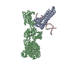
Entry Database : PDB / ID : 5dmqTitle Crystal structure of mouse eRF1 in complex with Reverse Transcriptase (RT) of Moloney Murine Leukemia Virus Eukaryotic peptide chain release factor subunit 1 Reverse transcriptase/ribonuclease H p80 Keywords / / / / / Function / homology Function Domain/homology Component
/ / / / / / / / / / / / / / / / / / / / / / / / / / / / / / / / / / / / / / / / / / / / / / / / / / / / / / / / / / / / / / / / / / / / / / / / / / / / / / / / / / / / / / / / / / / / / / / / / / / / / / / / / / / / / / / / / / / / / / / / / / / / Biological species Mus musculus (house mouse)Method / / Resolution : 4 Å Authors Tang, T. / Song, H. Journal : Nat Commun / Year : 2016Title : Structural basis of suppression of host translation termination by Moloney Murine Leukemia VirusAuthors : Tang, X. / Zhu, Y. / Baker, S.L. / Bowler, M.W. / Chen, B.J. / Chen, C. / Hogg, J.R. / Goff, S.P. / Song, H. History Deposition Sep 9, 2015 Deposition site / Processing site Revision 1.0 Jul 6, 2016 Provider / Type Revision 1.1 Oct 4, 2017 Group / Derived calculations / Category / pdbx_struct_oper_listItem / _pdbx_struct_oper_list.symmetry_operationRevision 1.2 Mar 20, 2024 Group / Database references / Category / chem_comp_bond / database_2Item / _database_2.pdbx_database_accession
Show all Show less
 Movie
Movie Controller

 Yorodumi
 Open data
 Basic information
 Components
 Keywords
 TRANSFERASE / HYDROLASE/TRANSLATION /
  Function and homology information

 Authors
 Citation
 Structure visualization
 Downloads & links
 Other downloads


 PDBj
PDBj
 Assembly
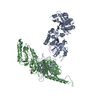

 Sample preparation
 / Beamline: X06SA / Wavelength: 1 Å
 Processing