 Movie
Movie Controller


+ Open data

- Basic information
| Entry | Database: PDB / ID: 5dkr | ||||||||||||
|---|---|---|---|---|---|---|---|---|---|---|---|---|---|
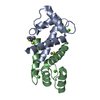
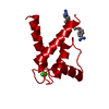
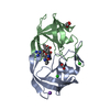
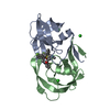
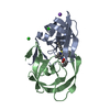
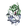
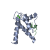
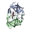
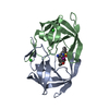
| Title | Crystal Structure of Calcium-loaded S100B bound to SBi29 | ||||||||||||
 Components Components | Protein S100-B | ||||||||||||
 Keywords Keywords | METAL BINDING PROTEIN/INHIBITOR /  malignant melanoma / calcium binding / complex / covalent inhibitor / METAL BINDING PROTEIN-INHIBITOR complex malignant melanoma / calcium binding / complex / covalent inhibitor / METAL BINDING PROTEIN-INHIBITOR complex | ||||||||||||
| Function / homology |  Function and homology information Function and homology informationAdvanced glycosylation endproduct receptor signaling / TRAF6 mediated NF-kB activation / TAK1-dependent IKK and NF-kappa-B activation / adaptive thermogenesis / kinase inhibitor activity / sympathetic neuron projection extension / positive regulation of complement activation / RAGE receptor binding / negative regulation of monocyte chemotactic protein-1 production / S100 protein binding ...Advanced glycosylation endproduct receptor signaling / TRAF6 mediated NF-kB activation / TAK1-dependent IKK and NF-kappa-B activation / adaptive thermogenesis / kinase inhibitor activity / sympathetic neuron projection extension / positive regulation of complement activation / RAGE receptor binding / negative regulation of monocyte chemotactic protein-1 production / S100 protein binding / axonogenesis / astrocyte activation / tau protein binding / calcium-dependent protein binding / regulation of translation / positive regulation of canonical NF-kappaB signal transduction / learning or memory / cell adhesion / phosphorylation / calcium ion binding / positive regulation of cell population proliferation / protein homodimerization activity / extracellular space / zinc ion binding / extracellular region / nucleus / cytoplasmSimilarity search - Function | ||||||||||||
| Biological species |  Bos taurus (cattle) Bos taurus (cattle) | ||||||||||||
| Method | X-RAY DIFFRACTION / SYNCHROTRON / MOLECULAR REPLACEMENT / Resolution: 1.742 Å | ||||||||||||
 Authors Authors | Cavalier, M.C. / Ansari, M.I. / Pierce, A.D. / Wilder, P.T. / McKnight, L.E. / Raman, E.P. / Neau, D.B. / Bezawada, P. / Alasady, M.J. / Varney, K.M. ...Cavalier, M.C. / Ansari, M.I. / Pierce, A.D. / Wilder, P.T. / McKnight, L.E. / Raman, E.P. / Neau, D.B. / Bezawada, P. / Alasady, M.J. / Varney, K.M. / Toth, E.A. / MacKerell Jr., A.D. / Coop, A. / Weber, D.J. | ||||||||||||
| Funding support |  United States, 3items United States, 3items
| ||||||||||||
 Citation Citation | Journal: J.Med.Chem. / Year: 2016 Title: Small Molecule Inhibitors of Ca(2+)-S100B Reveal Two Protein Conformations. Authors: Cavalier, M.C. / Ansari, M.I. / Pierce, A.D. / Wilder, P.T. / McKnight, L.E. / Raman, E.P. / Neau, D.B. / Bezawada, P. / Alasady, M.J. / Charpentier, T.H. / Varney, K.M. / Toth, E.A. / ...Authors: Cavalier, M.C. / Ansari, M.I. / Pierce, A.D. / Wilder, P.T. / McKnight, L.E. / Raman, E.P. / Neau, D.B. / Bezawada, P. / Alasady, M.J. / Charpentier, T.H. / Varney, K.M. / Toth, E.A. / MacKerell, A.D. / Coop, A. / Weber, D.J. | ||||||||||||
| History |
|
- Structure visualization
| Structure viewer | Molecule: MolmilJmol/JSmol |
|---|
- Downloads & links
-Download
| PDBx/mmCIF format | 5dkr.cif.gz | 58.5 KB | Display | PDBx/mmCIF format |
|---|---|---|---|---|
| PDB format | pdb5dkr.ent.gz | 40 KB | Display | PDB format |
| PDBx/mmJSON format | 5dkr.json.gz | Tree view | PDBx/mmJSON format | |
| Others |  Other downloads Other downloads |
-Validation report
| Arichive directory | https://data.pdbj.org/pub/pdb/validation_reports/dk/5dkrftp://data.pdbj.org/pub/pdb/validation_reports/dk/5dkr | HTTPS FTP |
|---|
-Related structure data
| Related structure data | 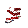 5dknC 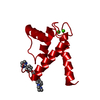 5dkqC  1mhoS C: citing same article ( S: Starting model for refinement |
|---|---|
| Similar structure data |

-Links
 PDBj
PDBj- Assembly
| Deposited unit | 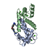
| ||||||||
|---|---|---|---|---|---|---|---|---|---|
| 1 |
| ||||||||
| Unit cell |
|
-Components
| #1: Protein | Mass: 10681.974 Da / Num. of mol.: 2 Source method: isolated from a genetically manipulated source Source: (gene. exp.) Bos taurus (cattle) / Gene: S100B / Production host:  Escherichia coli (E. coli) / References: UniProt: P02638 Escherichia coli (E. coli) / References: UniProt: P02638#2: Chemical | ChemComp-CA /   Mass: 40.078 Da / Num. of mol.: 5 / Source method: obtained synthetically / Formula: Ca Mass: 40.078 Da / Num. of mol.: 5 / Source method: obtained synthetically / Formula: Ca#3: Chemical | ChemComp-5CZ / |   Mass: 369.419 Da / Num. of mol.: 1 / Source method: obtained synthetically / Formula: C22H19N5O Mass: 369.419 Da / Num. of mol.: 1 / Source method: obtained synthetically / Formula: C22H19N5O#4: Water | ChemComp-HOH / | Water Mass: 18.015 Da / Num. of mol.: 172 / Source method: isolated from a natural source / Formula: H2O Mass: 18.015 Da / Num. of mol.: 172 / Source method: isolated from a natural source / Formula: H2O |
|---|
-Experimental details
-Experiment
| Experiment | Method: X-RAY DIFFRACTION / Number of used crystals: 1 |
|---|
- Sample preparation
| Crystal | Density Matthews: 2.34 Å3/Da / Density % sol: 47.54 % |
|---|---|
| Crystal grow | Temperature: 295 K / Method: vapor diffusion, sitting drop Details: 40% 2-methyl-2,4-pentanediol; 0.1M Hepes, pH 7.0, 7.5mM CaCl2 Temp details: 7 |
-Data collection
| Diffraction | Mean temperature: 100 K | |||||||||||||||||||||||||||
|---|---|---|---|---|---|---|---|---|---|---|---|---|---|---|---|---|---|---|---|---|---|---|---|---|---|---|---|---|
| Diffraction source | Source: SYNCHROTRON / Site: SSRL / Beamline: BL7-1 / Wavelength: 1.127092 Å | |||||||||||||||||||||||||||
| Detector | Type: ADSC QUANTUM 315r / Detector: CCD / Date: Dec 14, 2013 | |||||||||||||||||||||||||||
| Radiation | Protocol: SINGLE WAVELENGTH / Monochromatic (M) / Laue (L): M / Scattering type: x-ray | |||||||||||||||||||||||||||
| Radiation wavelength | Wavelength: 1.127092 Å / Relative weight: 1 | |||||||||||||||||||||||||||
| Reflection | Resolution: 1.74→33.3 Å / Num. obs: 20972 / % possible obs: 98.7 % / Redundancy: 3.9 % / Biso Wilson estimate: 28.49 Å2 / CC1/2: 0.997 / Rmerge(I) obs: 0.059 / Rpim(I) all: 0.033 / Net I/σ(I): 11.8 / Num. measured all: 82661 | |||||||||||||||||||||||||||
| Reflection shell | Diffraction-ID: 1 / Rejects: 0
|
- Processing
| Software |
| |||||||||||||||||||||||||||||||||||||||||||||||||||||||||||||||||||||||||||||||||||||||||||||||||||||||||
|---|---|---|---|---|---|---|---|---|---|---|---|---|---|---|---|---|---|---|---|---|---|---|---|---|---|---|---|---|---|---|---|---|---|---|---|---|---|---|---|---|---|---|---|---|---|---|---|---|---|---|---|---|---|---|---|---|---|---|---|---|---|---|---|---|---|---|---|---|---|---|---|---|---|---|---|---|---|---|---|---|---|---|---|---|---|---|---|---|---|---|---|---|---|---|---|---|---|---|---|---|---|---|---|---|---|---|
| Refinement | Method to determine structure: MOLECULAR REPLACEMENT Starting model: 1MHO Resolution: 1.742→33.298 Å / FOM work R set: 0.7799 / SU ML: 0.24 / Cross valid method: FREE R-VALUE / σ(F): 1.34 / Phase error: 28.12 / Stereochemistry target values: ML
| |||||||||||||||||||||||||||||||||||||||||||||||||||||||||||||||||||||||||||||||||||||||||||||||||||||||||
| Solvent computation | Shrinkage radii: 0.9 Å / VDW probe radii: 1.11 Å / Solvent model: FLAT BULK SOLVENT MODEL | |||||||||||||||||||||||||||||||||||||||||||||||||||||||||||||||||||||||||||||||||||||||||||||||||||||||||
| Displacement parameters | Biso max: 63.04 Å2 / Biso mean: 30.22 Å2 / Biso min: 14.5 Å2 | |||||||||||||||||||||||||||||||||||||||||||||||||||||||||||||||||||||||||||||||||||||||||||||||||||||||||
| Refinement step | Cycle: final / Resolution: 1.742→33.298 Å
| |||||||||||||||||||||||||||||||||||||||||||||||||||||||||||||||||||||||||||||||||||||||||||||||||||||||||
| Refine LS restraints |
| |||||||||||||||||||||||||||||||||||||||||||||||||||||||||||||||||||||||||||||||||||||||||||||||||||||||||
| LS refinement shell | Refine-ID: X-RAY DIFFRACTION / Total num. of bins used: 14
|
