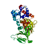
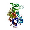
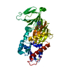
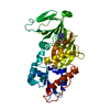
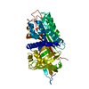
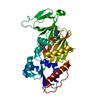
Entry Database : PDB / ID : 5dgiTitle Crystal structure of the catalytic domain of human diphosphoinositol pentakisphosphate kinase 2 (PPIP5K2) in complex with ADP and 3,5-(PCP)2-IP4 Inositol hexakisphosphate and diphosphoinositol-pentakisphosphate kinase 2 Keywords / Function / homology Function Domain/homology Component
/ / / / / / / / / / / / / / / / / / / / / / / / / / / / / / / / / / / / / / / / / / / / / / / / / / Biological species Homo sapiens (human)Method / / / Resolution : 1.85 Å Authors Wang, H. / Shears, S.B. Funding support Organization Grant number Country National Institutes of Health/National Institute of Environmental Health Sciences (NIH/NIEHS)
Journal : Chemistry / Year : 2016Title : Cellular Cations Control Conformational Switching of Inositol Pyrophosphate Analogues.Authors : Hager, A. / Wu, M. / Wang, H. / Brown, N.W. / Shears, S.B. / Veiga, N. / Fiedler, D. History Deposition Aug 27, 2015 Deposition site / Processing site Revision 1.0 Aug 10, 2016 Provider / Type Revision 1.1 Aug 24, 2016 Group Revision 1.2 Sep 20, 2017 Group / Derived calculations / Refinement descriptionCategory / pdbx_struct_oper_list / softwareItem / _pdbx_struct_oper_list.symmetry_operationRevision 1.3 Dec 18, 2019 Group / Category / Item Revision 1.4 Sep 27, 2023 Group / Database references / Refinement descriptionCategory chem_comp_atom / chem_comp_bond ... chem_comp_atom / chem_comp_bond / database_2 / pdbx_initial_refinement_model Item / _database_2.pdbx_database_accession
Show all Show less
 Movie
Movie Controller

 Yorodumi
 Open data
 Basic information
 Components
 Keywords
 TRANSFERASE / TRANSFERASE Inositol diphosphoinositol pentakisphosphate kinase Analog methylenebisphosphonate PPIP5K ATP-grasp Pyrophosphate Diphosphate
 Function and homology information
 Authors
 United States, 1items
  Citation
 Structure visualization
 Downloads & links
 Other downloads


 PDBj
PDBj


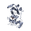
 Assembly


 Mass: 427.201 Da / Num. of mol.: 1 / Source method: obtained synthetically / Formula: C10H15N5O10P2 / Comment: ADP, energy-carrying molecule*YM
 Mass: 816.049 Da / Num. of mol.: 1 / Source method: obtained synthetically / Formula: C8H24O28P8
 Mass: 24.305 Da / Num. of mol.: 3 / Source method: obtained synthetically / Formula: Mg
 Mass: 59.044 Da / Num. of mol.: 3 / Source method: obtained synthetically / Formula: C2H3O2
 Mass: 62.068 Da / Num. of mol.: 5 / Source method: obtained synthetically / Formula: C2H6O2
 Sample preparation
 Processing