 Movie
Movie Controller

[English] 日本語
 Yorodumi
- PDB-5ddo: Structural and Dynamic Basis for Low Affinity-High Selectivity Bi... -
+ Open data

- Basic information
| Entry | Database: PDB / ID: 5ddo | ||||||
|---|---|---|---|---|---|---|---|
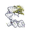
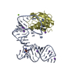
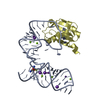
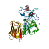
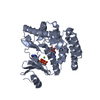
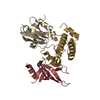
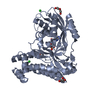
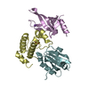
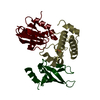
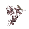
| Title | Structural and Dynamic Basis for Low Affinity-High Selectivity Binding of L-glutamine by the Gln-riboswitch | ||||||
 Components Components |
| ||||||
 Keywords Keywords | RNA BINDING PROTEIN/RNA /  riboswitch / L-glutamine / free-form / RNA / RNA BINDING PROTEIN-RNA complex riboswitch / L-glutamine / free-form / RNA / RNA BINDING PROTEIN-RNA complex | ||||||
| Function / homology |  Function and homology information Function and homology informationU1 snRNP binding / U1 snRNP / U1 snRNA binding / U4/U6 x U5 tri-snRNP complex / mRNA Splicing - Major Pathway / spliceosomal complex / mRNA splicing, via spliceosome / DNA binding / RNA binding / nucleoplasm ...U1 snRNP binding / U1 snRNP / U1 snRNA binding / U4/U6 x U5 tri-snRNP complex / mRNA Splicing - Major Pathway / spliceosomal complex / mRNA splicing, via spliceosome / DNA binding / RNA binding / nucleoplasm / identical protein binding / nucleusSimilarity search - Function | ||||||
| Biological species |  Homo sapiens (human) Homo sapiens (human) Synechococcus elongatus (bacteria) Synechococcus elongatus (bacteria) | ||||||
| Method | X-RAY DIFFRACTION / SYNCHROTRON / MOLECULAR REPLACEMENT / Resolution: 3.1 Å | ||||||
 Authors Authors | Ren, A. / Patel, D. | ||||||
| Funding support |  United States, 1items United States, 1items
| ||||||
 Citation Citation | Journal: Cell Rep / Year: 2015 Title: Structural and Dynamic Basis for Low-Affinity, High-Selectivity Binding of L-Glutamine by the Glutamine Riboswitch. Authors: Ren, A. / Xue, Y. / Peselis, A. / Serganov, A. / Al-Hashimi, H.M. / Patel, D.J. | ||||||
| History |
|
- Structure visualization
| Structure viewer | Molecule: MolmilJmol/JSmol |
|---|
- Downloads & links
-Download
| PDBx/mmCIF format | 5ddo.cif.gz | 111.8 KB | Display | PDBx/mmCIF format |
|---|---|---|---|---|
| PDB format | pdb5ddo.ent.gz | 81.2 KB | Display | PDB format |
| PDBx/mmJSON format | 5ddo.json.gz | Tree view | PDBx/mmJSON format | |
| Others |  Other downloads Other downloads |
-Validation report
| Arichive directory | https://data.pdbj.org/pub/pdb/validation_reports/dd/5ddoftp://data.pdbj.org/pub/pdb/validation_reports/dd/5ddo | HTTPS FTP |
|---|
-Related structure data


-Links
 PDBj
PDBj


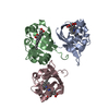
- Assembly
| Deposited unit | 
| ||||||||
|---|---|---|---|---|---|---|---|---|---|
| 1 | 
| ||||||||
| 2 | 
| ||||||||
| Unit cell |
|
-Components
| #1: RNA chain | Mass: 19718.770 Da / Num. of mol.: 2 / Source method: isolated from a natural source / Details: some nucleotides are too flexible in the structure / Source: (natural) Synechococcus elongatus (bacteria)#2: Protein | Mass: 11209.120 Da / Num. of mol.: 2 Source method: isolated from a genetically manipulated source Source: (gene. exp.) Homo sapiens (human) / Gene: SNRPA / Production host: Escherichia coli (E. coli) / References: UniProt: P09012#3: Water | ChemComp-HOH / | Water Mass: 18.015 Da / Num. of mol.: 11 / Source method: isolated from a natural source / Formula: H2O Mass: 18.015 Da / Num. of mol.: 11 / Source method: isolated from a natural source / Formula: H2O |
|---|
-Experimental details
-Experiment
| Experiment | Method: X-RAY DIFFRACTION |
|---|
- Sample preparation
| Crystal | Density Matthews: 2.84 Å3/Da / Density % sol: 56.76 % |
|---|---|
| Crystal grow | Temperature: 293 K / Method: evaporation / Details: 0.2 M Na-formate, 21% (w/v) PEG3350 |
-Data collection
| Diffraction | Mean temperature: 100 K |
|---|---|
| Diffraction source | Source: SYNCHROTRON / Site: APS / Beamline: 24-ID-E / Wavelength: 0.9792 Å |
| Detector | Type: ADSC QUANTUM 315 / Detector: CCD / Date: Jan 1, 2013 |
| Radiation | Protocol: SINGLE WAVELENGTH / Monochromatic (M) / Laue (L): M / Scattering type: x-ray |
| Radiation wavelength | Wavelength: 0.9792 Å / Relative weight: 1 |
| Reflection | Resolution: 3.1→50 Å / Num. obs: 12813 / % possible obs: 99.7 % / Redundancy: 4.1 % / Rmerge(I) obs: 0.088 / Net I/av σ(I): 15.7 / Net I/σ(I): 15.7 |
| Reflection shell | Resolution: 3.1→3.21 Å / Redundancy: 2.5 % / Rmerge(I) obs: 0.479 / Mean I/σ(I) obs: 2.5 / % possible all: 99.5 |
- Processing
| Software |
| |||||||||||||||||||||||||||||||||||
|---|---|---|---|---|---|---|---|---|---|---|---|---|---|---|---|---|---|---|---|---|---|---|---|---|---|---|---|---|---|---|---|---|---|---|---|---|
| Refinement | Method to determine structure: MOLECULAR REPLACEMENT Starting model: U1A protein Resolution: 3.1→39.917 Å / SU ML: 0.45 / Cross valid method: FREE R-VALUE / σ(F): 1.34 / Phase error: 31.11 / Stereochemistry target values: ML
| |||||||||||||||||||||||||||||||||||
| Solvent computation | Shrinkage radii: 1.24 Å / VDW probe radii: 1.4 Å / Solvent model: FLAT BULK SOLVENT MODEL / Bsol: 41.469 Å2 / ksol: 0.241 e/Å3 | |||||||||||||||||||||||||||||||||||
| Displacement parameters |
| |||||||||||||||||||||||||||||||||||
| Refinement step | Cycle: LAST / Resolution: 3.1→39.917 Å
| |||||||||||||||||||||||||||||||||||
| Refine LS restraints |
| |||||||||||||||||||||||||||||||||||
| LS refinement shell |
|
