 Movie
Movie Controller


+ Open data

- Basic information
| Entry | Database: PDB / ID: 5dbk | ||||||
|---|---|---|---|---|---|---|---|
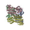
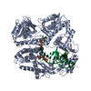
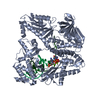
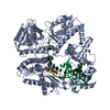
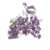
| Title | apo form of the quorum sensor NprR from B. thuringiensis | ||||||
 Components Components | Transcriptional regulator/TPR domain protein | ||||||
 Keywords Keywords |  SIGNALING PROTEIN / truncated form / TPR domain / mutant protein / asymmetric dimer SIGNALING PROTEIN / truncated form / TPR domain / mutant protein / asymmetric dimer | ||||||
| Function / homology |  Function and homology information Function and homology information | ||||||
| Biological species |  Bacillus thuringiensis Bt407 (bacteria) Bacillus thuringiensis Bt407 (bacteria) | ||||||
| Method | X-RAY DIFFRACTION / SYNCHROTRON / SAD / Resolution: 3.241 Å | ||||||
 Authors Authors | Talagas, A. / Perchat, S. / Lereclus, D. / Nessler, S. | ||||||
| Funding support |  France, 1items France, 1items
| ||||||
 Citation Citation | Journal: Plos Pathog. / Year: 2016 Title: How Quorum Sensing Connects Sporulation to Necrotrophism in Bacillus thuringiensis. Authors: Perchat, S. / Talagas, A. / Poncet, S. / Lazar, N. / Li de la Sierra-Gallay, I. / Gohar, M. / Lereclus, D. / Nessler, S. | ||||||
| History |
|
- Structure visualization
| Structure viewer | Molecule: MolmilJmol/JSmol |
|---|
- Downloads & links
-Download
| PDBx/mmCIF format | 5dbk.cif.gz | 152.8 KB | Display | PDBx/mmCIF format |
|---|---|---|---|---|
| PDB format | pdb5dbk.ent.gz | 121.1 KB | Display | PDB format |
| PDBx/mmJSON format | 5dbk.json.gz | Tree view | PDBx/mmJSON format | |
| Others |  Other downloads Other downloads |
-Validation report
| Arichive directory | https://data.pdbj.org/pub/pdb/validation_reports/db/5dbkftp://data.pdbj.org/pub/pdb/validation_reports/db/5dbk | HTTPS FTP |
|---|
-Related structure data
| Related structure data | |
|---|---|
| Similar structure data |


-Links
 PDBj
PDBj


- Assembly
| Deposited unit | 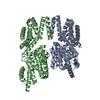
| ||||||||
|---|---|---|---|---|---|---|---|---|---|
| 1 |
| ||||||||
| Unit cell |
|
-Components
| #1: Protein | Mass: 44363.031 Da / Num. of mol.: 2 / Fragment: RESIDUES 13-375 / Mutation: Y223A, F225A Source method: isolated from a genetically manipulated source Details: THIS IS A TRUNCATED FORM OF THE PROTEIN, MISSING THE 60 RESIDUES OF THE N-TERMINAL HTH DOMAIN, WITH A C-TERMINAL HIS-TAG Source: (gene. exp.) Bacillus thuringiensis Bt407 (bacteria)Gene: nprR / Plasmid: pQE60 / Production host: Escherichia coli (E. coli) / Variant (production host): M15 [pRep4] / References: UniProt: M1Q3P9 |
|---|
-Experimental details
-Experiment
| Experiment | Method: X-RAY DIFFRACTION |
|---|
- Sample preparation
| Crystal | Density Matthews: 3.55 Å3/Da / Density % sol: 65.37 % |
|---|---|
| Crystal grow | Temperature: 291 K / Method: vapor diffusion / pH: 7.6 Details: NA CITRATE 1.0 M, HEPES 100 MM, PH 7.6, VAPOR DIFFUSION, TEMPERATURE 291K PH range: 7.6 |
-Data collection
| Diffraction | Mean temperature: 100 K |
|---|---|
| Diffraction source | Source: SYNCHROTRON / Site: ESRF / Beamline: ID29 / Wavelength: 0.976 Å |
| Detector | Type: DECTRIS PILATUS 6M-F / Detector: PIXEL / Date: Oct 25, 2013 |
| Radiation | Monochromator: SI(111) CRYSTAL / Protocol: SINGLE WAVELENGTH / Monochromatic (M) / Laue (L): M / Scattering type: x-ray |
| Radiation wavelength | Wavelength: 0.976 Å / Relative weight: 1 |
| Reflection | Resolution: 3.24→50 Å / Num. obs: 20499 / % possible obs: 98.4 % / Observed criterion σ(I): -3 / Redundancy: 3.46 % / Rmerge(I) obs: 0.12 / Rsym value: 0.12 / Net I/σ(I): 10.03 |
| Reflection shell | Resolution: 3.24→3.44 Å / Redundancy: 3.53 % / Rmerge(I) obs: 0.67 / Mean I/σ(I) obs: 2.18 / % possible all: 95.8 |
- Processing
| Software |
| ||||||||||||||||||||||||||||||||||||||||||||||||||||||||
|---|---|---|---|---|---|---|---|---|---|---|---|---|---|---|---|---|---|---|---|---|---|---|---|---|---|---|---|---|---|---|---|---|---|---|---|---|---|---|---|---|---|---|---|---|---|---|---|---|---|---|---|---|---|---|---|---|---|
| Refinement | Method to determine structure: SAD / Resolution: 3.241→47.929 Å / SU ML: 0.42 / Cross valid method: FREE R-VALUE / σ(F): 1.37 / Phase error: 26.96 / Stereochemistry target values: ML
| ||||||||||||||||||||||||||||||||||||||||||||||||||||||||
| Solvent computation | Shrinkage radii: 0.9 Å / VDW probe radii: 1.11 Å / Solvent model: FLAT BULK SOLVENT MODEL | ||||||||||||||||||||||||||||||||||||||||||||||||||||||||
| Refinement step | Cycle: LAST / Resolution: 3.241→47.929 Å
| ||||||||||||||||||||||||||||||||||||||||||||||||||||||||
| Refine LS restraints |
| ||||||||||||||||||||||||||||||||||||||||||||||||||||||||
| LS refinement shell |
|
