 Movie
Movie Controller


+ Open data

- Basic information
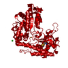
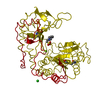
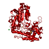
| Entry | Database: PDB / ID: 5d8g | ||||||
|---|---|---|---|---|---|---|---|
| Title | A structural view on the dissociation of E. coli Tryptophanase | ||||||
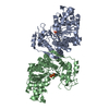
 Components Components | Tryptophanase | ||||||
 Keywords Keywords | LYASE / tryptophanase / PLP-dependent enzyme / cold dissociation / hydrophobic interactions / open conformation / closed conformation | ||||||
| Function / homology |  Function and homology information Function and homology informationindole metabolic process / tryptophanase activity / tryptophanase / cell pole / L-cysteine desulfhydrase activity / tryptophan catabolic process / potassium ion binding / pyridoxal phosphate binding / protein-containing complex / membrane ...indole metabolic process / tryptophanase activity / tryptophanase / cell pole / L-cysteine desulfhydrase activity / tryptophan catabolic process / potassium ion binding / pyridoxal phosphate binding / protein-containing complex / membrane / identical protein binding / cytosolSimilarity search - Function | ||||||
| Biological species |  Escherichia coli (E. coli) Escherichia coli (E. coli) | ||||||
| Method | X-RAY DIFFRACTION / MOLECULAR REPLACEMENT / Resolution: 1.89 Å | ||||||
 Authors Authors | Almog, O. | ||||||
 Citation Citation | Journal: Acta Crystallogr.,Sect.D / Year: 2015 Title: A structural view of the dissociation of Escherichia coli tryptophanase. Authors: Green, K. / Qasim, N. / Gdaelvsky, G. / Kogan, A. / Goldgur, Y. / Parola, A.H. / Lotan, O. / Almog, O. | ||||||
| History |
|
- Structure visualization
| Structure viewer | Molecule: MolmilJmol/JSmol |
|---|
- Downloads & links
-Download
| PDBx/mmCIF format | 5d8g.cif.gz | 114.7 KB | Display | PDBx/mmCIF format |
|---|---|---|---|---|
| PDB format | pdb5d8g.ent.gz | 92.5 KB | Display | PDB format |
| PDBx/mmJSON format | 5d8g.json.gz | Tree view | PDBx/mmJSON format | |
| Others |  Other downloads Other downloads |
-Validation report
| Arichive directory | https://data.pdbj.org/pub/pdb/validation_reports/d8/5d8gftp://data.pdbj.org/pub/pdb/validation_reports/d8/5d8g | HTTPS FTP |
|---|
-Related structure data
| Related structure data | |
|---|---|
| Similar structure data |

-Links
 PDBj
PDBj- Assembly
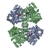
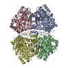
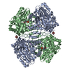
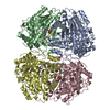
| Deposited unit | 
| ||||||||
|---|---|---|---|---|---|---|---|---|---|
| 1 | 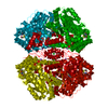
| ||||||||
| Unit cell |
| ||||||||
| Components on special symmetry positions |
|
-Components
-Protein , 1 types, 1 molecules A
| #1: Protein | / L-tryptophan indole-lyase / TNase Mass: 52496.992 Da / Num. of mol.: 1 Source method: isolated from a genetically manipulated source Details: HLPEPFRIR VIEPVKRTTR AYREEAIIKS GMNPFLLDS EDVFIDLLTD SGTGAMTQS MQAAMMRGDEAYSGSRSYYALA ESVKNIFGYQYTIPTHQGRGAEQIYIPVLIKKREQEKGLDRSKMVAFSNYFFDTTQGHSQINGCTVRNVYIKEAFDTGVR ...Details: HLPEPFRIR VIEPVKRTTR AYREEAIIKS GMNPFLLDS EDVFIDLLTD SGTGAMTQS MQAAMMRGDEAYSGSRSYYALA ESVKNIFGYQYTIPTHQGRGAEQIYIPVLIKKREQEKGLDRSKMVAFSNYFFDTTQGHSQINGCTVRNVYIKEAFDTGVR YDFKGNFDLEGLERGIEEVGPNNVPYIVATITSNSAGGQPVSLANLKAMYSIAKKYDIPVVMDSARFAENAYFIKQREAE YKDWTIEQITRETYKYADMLAMSAKKDAMVPMGGLLCMKDDSFFDVYTECRTLCVVQEGFPTYGGLEGGAMERLAVGLYD GMNLDWLAYRIAQVQYLVDGLEEIGVVCQQAGGHAAFVDAGKLLPHIPADQFPAQALACELYKVAGIRAVEIGSFLLGRD PKTGKQLPCPAELLRLTIPRATYTQTHMDFIIEAFKHVKENAANIKGLTFTYEPKVLRHFTAKLKEV Source: (gene. exp.) Escherichia coli (strain K12) (bacteria)Strain: K12 / Gene: tnaA, ind, b3708, JW3686 Production host: Escherichia coli str. 'clone D i2' (bacteria)References: UniProt: P0A853, tryptophanase |
|---|
-Non-polymers , 6 types, 429 molecules 


| #2: Chemical | ChemComp-CL / Chloride Mass: 35.453 Da / Num. of mol.: 1 / Source method: obtained synthetically / Formula: Cl Mass: 35.453 Da / Num. of mol.: 1 / Source method: obtained synthetically / Formula: Cl | ||||||||
|---|---|---|---|---|---|---|---|---|---|
| #3: Chemical |  Mass: 24.305 Da / Num. of mol.: 3 / Source method: obtained synthetically / Formula: Mg Mass: 24.305 Da / Num. of mol.: 3 / Source method: obtained synthetically / Formula: Mg#4: Chemical | ChemComp-EPE / | HEPES Mass: 238.305 Da / Num. of mol.: 1 / Source method: obtained synthetically / Formula: C8H18N2O4S / Comment: pH buffer*YM Mass: 238.305 Da / Num. of mol.: 1 / Source method: obtained synthetically / Formula: C8H18N2O4S / Comment: pH buffer*YM#5: Chemical | ChemComp-NA / |  Mass: 22.990 Da / Num. of mol.: 1 / Source method: obtained synthetically / Formula: Na Mass: 22.990 Da / Num. of mol.: 1 / Source method: obtained synthetically / Formula: Na#6: Chemical | ChemComp-CA / |  Mass: 40.078 Da / Num. of mol.: 1 / Source method: obtained synthetically / Formula: Ca Mass: 40.078 Da / Num. of mol.: 1 / Source method: obtained synthetically / Formula: Ca#7: Water | ChemComp-HOH / | WaterMass: 18.015 Da / Num. of mol.: 422 / Source method: isolated from a natural source / Formula: H2O |
-Experimental details
-Experiment
| Experiment | Method: X-RAY DIFFRACTION |
|---|
- Sample preparation
| Crystal | Density Matthews: 2.92 Å3/Da / Density % sol: 57.83 % |
|---|---|
| Crystal grow | Temperature: 293 K / Method: vapor diffusion Details: 30%(w/v) PEG 400, 100 mM HEPES pH 7.5, 200 mM MgCl2, 5 mM 2-mercaptoethanol |
-Data collection
| Diffraction | Mean temperature: 100 K |
|---|---|
| Diffraction source | Source: ROTATING ANODE / Type: RIGAKU RU200 / Wavelength: 1.54 Å |
| Detector | Type: RIGAKU RAXIS IV / Detector: IMAGE PLATE / Date: Apr 14, 2010 |
| Radiation | Protocol: SINGLE WAVELENGTH / Monochromatic (M) / Laue (L): M / Scattering type: x-ray |
| Radiation wavelength | Wavelength: 1.54 Å / Relative weight: 1 |
| Reflection | Resolution: 1.89→49.2 Å / Num. obs: 45940 / % possible obs: 99 % / Redundancy: 6.6 % / Net I/σ(I): 10.2 |
| Reflection shell | Highest resolution: 1.89 Å |
- Processing
| Software |
| ||||||||||||||||||||||||||||||||||||||||||||||||||||||||||||||||||||||||||||||||||||||||||||||||||||||||||||||||||||||||||||||||||||||||||||||||||||||||||||||||||||||||||||||||||||||
|---|---|---|---|---|---|---|---|---|---|---|---|---|---|---|---|---|---|---|---|---|---|---|---|---|---|---|---|---|---|---|---|---|---|---|---|---|---|---|---|---|---|---|---|---|---|---|---|---|---|---|---|---|---|---|---|---|---|---|---|---|---|---|---|---|---|---|---|---|---|---|---|---|---|---|---|---|---|---|---|---|---|---|---|---|---|---|---|---|---|---|---|---|---|---|---|---|---|---|---|---|---|---|---|---|---|---|---|---|---|---|---|---|---|---|---|---|---|---|---|---|---|---|---|---|---|---|---|---|---|---|---|---|---|---|---|---|---|---|---|---|---|---|---|---|---|---|---|---|---|---|---|---|---|---|---|---|---|---|---|---|---|---|---|---|---|---|---|---|---|---|---|---|---|---|---|---|---|---|---|---|---|---|---|
| Refinement | Method to determine structure: MOLECULAR REPLACEMENT / Resolution: 1.89→49.2 Å / Cor.coef. Fo:Fc: 0.957 / Cor.coef. Fo:Fc free: 0.941 / SU B: 2.511 / SU ML: 0.076 / Cross valid method: THROUGHOUT / ESU R: 0.127 / ESU R Free: 0.124 / Stereochemistry target values: MAXIMUM LIKELIHOOD / Details: HYDROGENS HAVE BEEN ADDED IN THE RIDING POSITIONS
| ||||||||||||||||||||||||||||||||||||||||||||||||||||||||||||||||||||||||||||||||||||||||||||||||||||||||||||||||||||||||||||||||||||||||||||||||||||||||||||||||||||||||||||||||||||||
| Solvent computation | Ion probe radii: 0.8 Å / Shrinkage radii: 0.8 Å / VDW probe radii: 1.4 Å / Solvent model: MASK | ||||||||||||||||||||||||||||||||||||||||||||||||||||||||||||||||||||||||||||||||||||||||||||||||||||||||||||||||||||||||||||||||||||||||||||||||||||||||||||||||||||||||||||||||||||||
| Displacement parameters | Biso mean: 25.383 Å2
| ||||||||||||||||||||||||||||||||||||||||||||||||||||||||||||||||||||||||||||||||||||||||||||||||||||||||||||||||||||||||||||||||||||||||||||||||||||||||||||||||||||||||||||||||||||||
| Refinement step | Cycle: LAST / Resolution: 1.89→49.2 Å
| ||||||||||||||||||||||||||||||||||||||||||||||||||||||||||||||||||||||||||||||||||||||||||||||||||||||||||||||||||||||||||||||||||||||||||||||||||||||||||||||||||||||||||||||||||||||
| Refine LS restraints |
|