[English] 日本語
Yorodumi
- PDB-5d79: Structure of BBE-like #28 from Arabidopsis thaliana -

+
Open data


ID or keywords:

Loading...

-
Basic information

Entry
Database: PDB / ID: 5d79
TitleStructure of BBE-like #28 from Arabidopsis thaliana
ComponentsBerberine bridge enzyme-like protein
KeywordsOXIDOREDUCTASE / covalent FAD binding / berberine bridge enzyme like / plant enzyme
Function / homology
Function and homology information


cell wall / Oxidoreductases; Acting on the CH-OH group of donors; With NAD+ or NADP+ as acceptor / response to salt stress / FAD binding / oxidoreductase activity / extracellular region
Similarity search - Function
Vanillyl-alcohol Oxidase; Chain A, domain 3 - #20 / Berberine/berberine-like / Berberine and berberine like / Vanillyl-alcohol Oxidase; Chain A, domain 3 / Uridine Diphospho-n-acetylenolpyruvylglucosamine Reductase, domain 2 / Uridine Diphospho-n-acetylenolpyruvylglucosamine Reductase; domain 2 / Uridine Diphospho-n-acetylenolpyruvylglucosamine Reductase; domain 3 - #10 / Uridine Diphospho-n-acetylenolpyruvylglucosamine Reductase; domain 3 / FAD linked oxidase, N-terminal / FAD binding domain ...Vanillyl-alcohol Oxidase; Chain A, domain 3 - #20 / Berberine/berberine-like / Berberine and berberine like / Vanillyl-alcohol Oxidase; Chain A, domain 3 / Uridine Diphospho-n-acetylenolpyruvylglucosamine Reductase, domain 2 / Uridine Diphospho-n-acetylenolpyruvylglucosamine Reductase; domain 2 / Uridine Diphospho-n-acetylenolpyruvylglucosamine Reductase; domain 3 - #10 / Uridine Diphospho-n-acetylenolpyruvylglucosamine Reductase; domain 3 / FAD linked oxidase, N-terminal / FAD binding domain / FAD-binding, type PCMH, subdomain 1 / FAD-binding domain, PCMH-type / PCMH-type FAD-binding domain profile. / FAD-binding, type PCMH-like superfamily / 2-Layer Sandwich / 3-Layer(aba) Sandwich / Alpha Beta
Similarity search - Domain/homology
FLAVIN-ADENINE DINUCLEOTIDE / PHOSPHATE ION / Berberine bridge enzyme-like 28
Similarity search - Component
Biological speciesArabidopsis thaliana (thale cress)
MethodX-RAY DIFFRACTION / SYNCHROTRON / MOLECULAR REPLACEMENT / Resolution: 1.849 Å
AuthorsDaniel, B. / Kumar, P. / Gruber, K.
Funding support Austria, 1items
OrganizationGrant numberCountry
Austrian Science FundW901 Austria
CitationJournal: Plos One / Year: 2016
Title: Structure of a Berberine Bridge Enzyme-Like Enzyme with an Active Site Specific to the Plant Family Brassicaceae.
Authors: Daniel, B. / Wallner, S. / Steiner, B. / Oberdorfer, G. / Kumar, P. / van der Graaff, E. / Roitsch, T. / Sensen, C.W. / Gruber, K. / Macheroux, P.
History
DepositionAug 13, 2015Deposition site: RCSB / Processing site: PDBE
Revision 1.0Jun 22, 2016Provider: repository / Type: Initial release
Revision 1.1Sep 6, 2017Group: Advisory / Author supporting evidence / Derived calculations
Category: pdbx_audit_support / pdbx_validate_close_contact / struct_conn
Item: _pdbx_audit_support.funding_organization
Revision 2.0Jan 10, 2024Group: Atomic model / Data collection ...Atomic model / Data collection / Database references / Refinement description
Category: atom_site / chem_comp_atom ...atom_site / chem_comp_atom / chem_comp_bond / database_2 / pdbx_initial_refinement_model
Item: _atom_site.occupancy / _database_2.pdbx_DOI / _database_2.pdbx_database_accession

-
Structure visualization

Structure viewerMolecule:
MolmilJmol/JSmol

Downloads & links

-
Assembly

Deposited unit
A: Berberine bridge enzyme-like protein
B: Berberine bridge enzyme-like protein
hetero molecules


Theoretical massNumber of molelcules
Total (without water)122,2578
Polymers120,4252
Non-polymers1,8326
Water8,575476
1
A: Berberine bridge enzyme-like protein
hetero molecules


Theoretical massNumber of molelcules
Total (without water)61,1294
Polymers60,2131
Non-polymers9163
Water181
TypeNameSymmetry operationNumber
identity operation1_555x,y,z1
2
B: Berberine bridge enzyme-like protein
hetero molecules


Theoretical massNumber of molelcules
Total (without water)61,1294
Polymers60,2131
Non-polymers9163
Water181
TypeNameSymmetry operationNumber
identity operation1_555x,y,z1
Unit cell
Length a, b, c (Å)127.024, 133.132, 138.999
Angle α, β, γ (deg.)90.00, 90.00, 90.00
Int Tables number20
Space group name H-MC2221
Components on special symmetry positions
IDModelComponents
11A-921-

HOH

21A-966-

HOH

-
Components

#1: Protein Berberine bridge enzyme-like protein / FAD-binding and BBE domain-containing protein


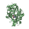
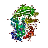
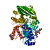
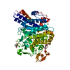
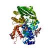
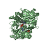
Mass: 60212.555 Da / Num. of mol.: 2
Source method: isolated from a genetically manipulated source
Details: THE FAD COFACTOR IS COVALENTLY LINKED TO THE PEPTIDE CHAIN VIA A COVALENT BOND BETWEEN ND1 OF HIS111 AND THE 8-ALPHA-METHYL GROUP OF THE ISOALLOXAZINE RING.
Source: (gene. exp.) Arabidopsis thaliana (thale cress) / Gene: At5g44440 / Plasmid: pPICZa / Production host: Komagataella pastoris (fungus) / Strain (production host): KM71H / References: UniProt: Q9FI21
#2: Chemical ChemComp-CL / CHLORIDE ION / Chloride


Mass: 35.453 Da / Num. of mol.: 2 / Source method: obtained synthetically / Formula: Cl
#3: Chemical ChemComp-PO4 / PHOSPHATE ION / Phosphate


Mass: 94.971 Da / Num. of mol.: 2 / Source method: obtained synthetically / Formula: PO4
#4: Chemical ChemComp-FAD / FLAVIN-ADENINE DINUCLEOTIDE / Flavin adenine dinucleotide


Mass: 785.550 Da / Num. of mol.: 2 / Source method: obtained synthetically / Formula: C27H33N9O15P2 / Comment: FAD*YM
#5: Water ChemComp-HOH / water / Water


Mass: 18.015 Da / Num. of mol.: 476 / Source method: isolated from a natural source / Formula: H2O

-
Experimental details

-
Experiment

ExperimentMethod: X-RAY DIFFRACTION

-
Sample preparation

CrystalDensity Matthews: 2.45 Å3/Da / Density % sol: 49.83 % / Description: tetragonal bipyramidal
Crystal growTemperature: 293 K / Method: microbatch / pH: 7
Details: 0.1 M HEPES buffer pH 7.0, 30 % v/v Jeffamine ED 2001

-
Data collection

DiffractionMean temperature: 100 K
Diffraction sourceSource: SYNCHROTRON / Site: ESRF / Beamline: BM14 / Wavelength: 0.91964 Å
DetectorType: MARMOSAIC 225 mm CCD / Detector: CCD / Date: Jun 20, 2013 / Details: bent collimating mirror and toroid
RadiationMonochromator: Si(111) monochromator / Protocol: SINGLE WAVELENGTH / Monochromatic (M) / Laue (L): M / Scattering type: x-ray
Radiation wavelengthWavelength: 0.91964 Å / Relative weight: 1
ReflectionResolution: 1.849→15.89 Å / Num. all: 93337 / Num. obs: 93337 / % possible obs: 93.16 % / Observed criterion σ(F): 0 / Observed criterion σ(I): 0 / Redundancy: 4 % / Biso Wilson estimate: 20.4 Å2 / Rmerge(I) obs: 0.1224 / Net I/σ(I): 7.22
Reflection shellResolution: 1.849→1.915 Å / Redundancy: 3.9 % / Rmerge(I) obs: 0.5576 / % possible all: 91.72

-
Processing

Software
NameVersionClassification
PHENIX1.8.3_1479refinement
XDSdata reduction
SCALAdata scaling
PHASERphasing
RefinementMethod to determine structure: MOLECULAR REPLACEMENT
Starting model: 3D2H
Resolution: 1.849→15.887 Å / Cross valid method: FREE R-VALUE / σ(F): 1.34 / Phase error: 26.43 / Stereochemistry target values: TWIN_LSQ_F
RfactorNum. reflection% reflectionSelection details
Rfree0.2429 4947 5.3 %random selection
Rwork0.1965 ---
obs0.1988 93334 93.16 %-
Solvent computationShrinkage radii: 0.9 Å / VDW probe radii: 1.11 Å / Solvent model: FLAT BULK SOLVENT MODEL
Refinement stepCycle: LAST / Resolution: 1.849→15.887 Å
ProteinNucleic acidLigandSolventTotal
Num. atoms7875 0 118 476 8469
Refine LS restraints
Refine-IDTypeDev idealNumber
X-RAY DIFFRACTIONf_bond_d0.0088199
X-RAY DIFFRACTIONf_angle_d1.0511106
X-RAY DIFFRACTIONf_dihedral_angle_d13.2783012
X-RAY DIFFRACTIONf_chiral_restr0.0391192
X-RAY DIFFRACTIONf_plane_restr0.0051399
LS refinement shell
Resolution (Å)Rfactor RfreeNum. reflection RfreeRfactor RworkNum. reflection RworkRefine-ID% reflection obs (%)
1.8515-1.88330.32912190.30654384X-RAY DIFFRACTION89
1.8833-1.91750.2922400.28994553X-RAY DIFFRACTION92
1.9175-1.95430.29822370.26394515X-RAY DIFFRACTION91
1.9543-1.99410.29232330.26334497X-RAY DIFFRACTION91
1.9941-2.03740.29222280.25784512X-RAY DIFFRACTION91
2.0374-2.08470.2842300.24864537X-RAY DIFFRACTION91
2.0847-2.13670.26392290.24134490X-RAY DIFFRACTION91
2.1367-2.19430.29762350.23864479X-RAY DIFFRACTION90
2.1943-2.25860.24712660.2364418X-RAY DIFFRACTION89
2.2586-2.33130.27792480.23344441X-RAY DIFFRACTION90
2.3313-2.41430.27922120.22574502X-RAY DIFFRACTION90
2.4143-2.51060.27462450.22794368X-RAY DIFFRACTION89
2.5106-2.62430.26122240.2224448X-RAY DIFFRACTION89
2.6243-2.76190.25742480.21684382X-RAY DIFFRACTION88
2.7619-2.93370.24762240.20654364X-RAY DIFFRACTION88
2.9337-3.15840.25442260.19714368X-RAY DIFFRACTION87
3.1584-3.47280.24412290.17854325X-RAY DIFFRACTION86
3.4728-3.96760.20772280.15094330X-RAY DIFFRACTION86
3.9676-4.96970.18872390.12344295X-RAY DIFFRACTION85
4.9697-15.22550.20122220.1534401X-RAY DIFFRACTION84
Refinement TLS params.

Method: refined / Refine-ID: X-RAY DIFFRACTION

IDL112)L122)L132)L222)L232)L332)S11 (Å °)S12 (Å °)S13 (Å °)S21 (Å °)S22 (Å °)S23 (Å °)S31 (Å °)S32 (Å °)S33 (Å °)T112)T122)T132)T222)T232)T332)Origin x (Å)Origin y (Å)Origin z (Å)
15.9271-1.77142.44336.4156-0.58241.08940.1123-0.3829-0.44360.1725-0.1208-0.1710.1769-0.0568-0.05540.49650.2950.06670.70920.23080.40898.7904-11.6586118.492
20.4238-0.3613-0.35481.17-0.45051.0007-0.1226-0.3229-0.11610.38330.0144-0.19120.18680.49280.09670.25380.11750.00450.46870.11140.213595.06331.9053109.3993
31.0101-0.12-0.50480.8071-0.1091.4716-0.04820.0236-0.1197-0.05210.00270.01050.08750.07640.03860.09770.01660.01370.1287-0.00580.116382.39558.66286.9382
40.89780.1124-0.43210.89240.0021.1196-0.0071-0.19450.03080.1199-0.0105-0.0289-0.04420.1548-0.00480.11660.01770.00620.1676-0.01160.123583.508918.6765100.9296
54.08655.10284.74297.65227.66937.88780.4557-0.96921.1972-0.06470.3574-0.3198-2.03480.0519-0.8091.0971-0.04240.5151.0334-0.27980.657669.858226.532249.3171
60.0241-0.01050.05060.2175-0.13330.77570.3330.9021-0.4888-0.1971-0.1858-0.30190.370.35050.04410.39810.2731-0.03440.6408-0.35010.356259.92131.594264.4621
72.1991-0.19720.63080.57230.12721.13820.11090.1936-0.1522-0.032-0.0340.02410.04980.0633-0.04480.16490.05490.0110.13590.00560.119252.066345.505984.1906
80.56120.0677-0.07940.4262-0.15341.9255-0.00650.6555-0.0473-0.1615-0.01630.01570.0016-0.1120.01890.23810.0965-0.00150.4510.00960.163945.875648.846863.5946
Refinement TLS group
IDRefine-IDRefine TLS-IDSelection details
1X-RAY DIFFRACTION1(chain A and resid 33:47)
2X-RAY DIFFRACTION2(chain A and resid 48:143)
3X-RAY DIFFRACTION3(chain A and resid 144:344)
4X-RAY DIFFRACTION4(chain A and resid 345:530)
5X-RAY DIFFRACTION5(chain B and resid 33:47)
6X-RAY DIFFRACTION6(chain B and resid 48:143)
7X-RAY DIFFRACTION7(chain B and resid 144:444)
8X-RAY DIFFRACTION8(chain B and resid 445:530)

+
About Yorodumi

-
News

-
Feb 9, 2022. New format data for meta-information of EMDB entries

New format data for meta-information of EMDB entries

  • Version 3 of the EMDB header file is now the official format.
  • The previous official version 1.9 will be removed from the archive.

Related info.:EMDB header

External links:wwPDB to switch to version 3 of the EMDB data model

-
Aug 12, 2020. Covid-19 info

Covid-19 info

URL: https://pdbj.org/emnavi/covid19.php

New page: Covid-19 featured information page in EM Navigator.

Related info.:Covid-19 info / Mar 5, 2020. Novel coronavirus structure data

+
Mar 5, 2020. Novel coronavirus structure data

Novel coronavirus structure data

Related info.:Yorodumi Speices / Aug 12, 2020. Covid-19 info

External links:COVID-19 featured content - PDBj / Molecule of the Month (242):Coronavirus Proteases

+
Jan 31, 2019. EMDB accession codes are about to change! (news from PDBe EMDB page)

EMDB accession codes are about to change! (news from PDBe EMDB page)

  • The allocation of 4 digits for EMDB accession codes will soon come to an end. Whilst these codes will remain in use, new EMDB accession codes will include an additional digit and will expand incrementally as the available range of codes is exhausted. The current 4-digit format prefixed with “EMD-” (i.e. EMD-XXXX) will advance to a 5-digit format (i.e. EMD-XXXXX), and so on. It is currently estimated that the 4-digit codes will be depleted around Spring 2019, at which point the 5-digit format will come into force.
  • The EM Navigator/Yorodumi systems omit the EMD- prefix.

Related info.:Q: What is EMD? / ID/Accession-code notation in Yorodumi/EM Navigator

External links:EMDB Accession Codes are Changing Soon! / Contact to PDBj

+
Jul 12, 2017. Major update of PDB

Major update of PDB

  • wwPDB released updated PDB data conforming to the new PDBx/mmCIF dictionary.
  • This is a major update changing the version number from 4 to 5, and with Remediation, in which all the entries are updated.
  • In this update, many items about electron microscopy experimental information are reorganized (e.g. em_software).
  • Now, EM Navigator and Yorodumi are based on the updated data.

External links:wwPDB Remediation / Enriched Model Files Conforming to OneDep Data Standards Now Available in the PDB FTP Archive

-
Yorodumi

Thousand views of thousand structures

  • Yorodumi is a browser for structure data from EMDB, PDB, SASBDB, etc.
  • This page is also the successor to EM Navigator detail page, and also detail information page/front-end page for Omokage search.
  • The word "yorodu" (or yorozu) is an old Japanese word meaning "ten thousand". "mi" (miru) is to see.

Related info.:EMDB / PDB / SASBDB / Comparison of 3 databanks / Yorodumi Search / Aug 31, 2016. New EM Navigator & Yorodumi / Yorodumi Papers / Jmol/JSmol / Function and homology information / Changes in new EM Navigator and Yorodumi

Read more