 Movie
Movie Controller


+ Open data

- Basic information
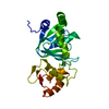
| Entry | Database: PDB / ID: 5d74 | ||||||
|---|---|---|---|---|---|---|---|
| Title | The crystal structure of Ly7917 | ||||||
 Components Components | Putative phage lysin | ||||||
 Keywords Keywords |  HYDROLASE / endolysin / prophage / Lytic activity HYDROLASE / endolysin / prophage / Lytic activity | ||||||
| Function / homology | Bacterial SH3 domain / SH3b domain profile. / Bacterial SH3 domain homologues / CHAP domain profile. / CHAP domain / CHAP domain / SH3-like domain, bacterial-type / Papain-like cysteine peptidase superfamily / Putative phage lysin Function and homology information Function and homology information | ||||||
| Biological species |  Streptococcus phage phi7917 (virus) Streptococcus phage phi7917 (virus) | ||||||
| Method | X-RAY DIFFRACTION / SYNCHROTRON / SAD / Resolution: 1.9 Å | ||||||
 Authors Authors | Wu, L. / Ji, W.H. / Sun, J.H. / Zhou, J.H. | ||||||
 Citation Citation | Journal: To Be Published Title: The crystal structure of Ly7917 Authors: Wu, L. / Ji, W.H. / Sun, J.H. / Zhou, J.H. | ||||||
| History |
|
- Structure visualization
| Structure viewer | Molecule: MolmilJmol/JSmol |
|---|
- Downloads & links
-Download
| PDBx/mmCIF format | 5d74.cif.gz | 115.4 KB | Display | PDBx/mmCIF format |
|---|---|---|---|---|
| PDB format | pdb5d74.ent.gz | 87.7 KB | Display | PDB format |
| PDBx/mmJSON format | 5d74.json.gz | Tree view | PDBx/mmJSON format | |
| Others |  Other downloads Other downloads |
-Validation report
| Arichive directory | https://data.pdbj.org/pub/pdb/validation_reports/d7/5d74ftp://data.pdbj.org/pub/pdb/validation_reports/d7/5d74 | HTTPS FTP |
|---|
-Related structure data


-Links
 PDBj
PDBj- Assembly
| Deposited unit | 
| ||||||||
|---|---|---|---|---|---|---|---|---|---|
| 1 | 
| ||||||||
| 2 | 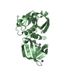
| ||||||||
| Unit cell |
| ||||||||
| Components on special symmetry positions |
|
-Components
| #1: Protein | Mass: 29354.111 Da / Num. of mol.: 2 Source method: isolated from a genetically manipulated source Source: (gene. exp.) Streptococcus phage phi7917 (virus) / Gene: phi7917_002 / Plasmid: pSJ2 / Production host:  Escherichia coli BL21(DE3) (bacteria) / Strain (production host): BL21(DE3) / References: UniProt: M1PKZ3 Escherichia coli BL21(DE3) (bacteria) / Strain (production host): BL21(DE3) / References: UniProt: M1PKZ3#2: Chemical | ChemComp-CL / Chloride  Mass: 35.453 Da / Num. of mol.: 6 / Source method: obtained synthetically / Formula: Cl Mass: 35.453 Da / Num. of mol.: 6 / Source method: obtained synthetically / Formula: Cl#3: Chemical | ChemComp-NA /   Mass: 22.990 Da / Num. of mol.: 7 / Source method: obtained synthetically / Formula: Na Mass: 22.990 Da / Num. of mol.: 7 / Source method: obtained synthetically / Formula: Na#4: Water | ChemComp-HOH / | Water Mass: 18.015 Da / Num. of mol.: 493 / Source method: isolated from a natural source / Formula: H2O Mass: 18.015 Da / Num. of mol.: 493 / Source method: isolated from a natural source / Formula: H2O |
|---|
-Experimental details
-Experiment
| Experiment | Method: X-RAY DIFFRACTION |
|---|
- Sample preparation
| Crystal | Density Matthews: 2.71 Å3/Da / Density % sol: 54.63 % |
|---|---|
| Crystal grow | Temperature: 291 K / Method: vapor diffusion, sitting drop / pH: 7 / Details: 0.1M Tris, 0.8M LiCl / PH range: 6.5-7.0 |
-Data collection
| Diffraction | Mean temperature: 100 K |
|---|---|
| Diffraction source | Source: SYNCHROTRON / Site: SSRF  / Beamline: BL19U1 / Wavelength: 0.97852 Å / Beamline: BL19U1 / Wavelength: 0.97852 Å |
| Detector | Type: PSI PILATUS 6M / Detector: PIXEL / Date: Dec 3, 2014 |
| Radiation | Protocol: SINGLE WAVELENGTH / Monochromatic (M) / Laue (L): M / Scattering type: x-ray |
| Radiation wavelength | Wavelength: 0.97852 Å / Relative weight: 1 |
| Reflection | Resolution: 1.9→50 Å / Num. obs: 47363 / % possible obs: 99.8 % / Redundancy: 13.1 % / Net I/σ(I): 15.9 |
| Reflection shell | Resolution: 1.9→1.97 Å / Redundancy: 11.9 % |
- Processing
| Software |
| ||||||||||||||||||||||||||||||||||||||||||||||||||||||||||||||||||||||||||||||||||||||||||||||||||||||||||||||||||||||||||||||
|---|---|---|---|---|---|---|---|---|---|---|---|---|---|---|---|---|---|---|---|---|---|---|---|---|---|---|---|---|---|---|---|---|---|---|---|---|---|---|---|---|---|---|---|---|---|---|---|---|---|---|---|---|---|---|---|---|---|---|---|---|---|---|---|---|---|---|---|---|---|---|---|---|---|---|---|---|---|---|---|---|---|---|---|---|---|---|---|---|---|---|---|---|---|---|---|---|---|---|---|---|---|---|---|---|---|---|---|---|---|---|---|---|---|---|---|---|---|---|---|---|---|---|---|---|---|---|---|
| Refinement | Method to determine structure: SAD / Resolution: 1.9→34.643 Å / SU ML: 0.22 / Cross valid method: FREE R-VALUE / σ(F): 1.34 / Phase error: 24.17 / Stereochemistry target values: ML
| ||||||||||||||||||||||||||||||||||||||||||||||||||||||||||||||||||||||||||||||||||||||||||||||||||||||||||||||||||||||||||||||
| Solvent computation | Shrinkage radii: 0.9 Å / VDW probe radii: 1.11 Å / Solvent model: FLAT BULK SOLVENT MODEL | ||||||||||||||||||||||||||||||||||||||||||||||||||||||||||||||||||||||||||||||||||||||||||||||||||||||||||||||||||||||||||||||
| Refinement step | Cycle: LAST / Resolution: 1.9→34.643 Å
| ||||||||||||||||||||||||||||||||||||||||||||||||||||||||||||||||||||||||||||||||||||||||||||||||||||||||||||||||||||||||||||||
| Refine LS restraints |
| ||||||||||||||||||||||||||||||||||||||||||||||||||||||||||||||||||||||||||||||||||||||||||||||||||||||||||||||||||||||||||||||
| LS refinement shell |
|
