 Movie
Movie Controller

[English] 日本語
 Yorodumi
- PDB-5crv: Crystal structure of the Bro domain of HD-PTP in a complex with t... -
+ Open data

- Basic information
| Entry | Database: PDB / ID: 5crv | ||||||
|---|---|---|---|---|---|---|---|
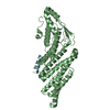
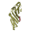
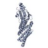
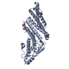
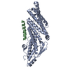
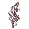
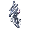
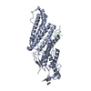
| Title | Crystal structure of the Bro domain of HD-PTP in a complex with the core region of STAM2 | ||||||
 Components Components |
| ||||||
 Keywords Keywords |  PROTEIN TRANSPORT / HD-PTP / Bro domain / STAM2 / ESCRT-0 / EGFR PROTEIN TRANSPORT / HD-PTP / Bro domain / STAM2 / ESCRT-0 / EGFR | ||||||
| Function / homology |  Function and homology information Function and homology informationpositive regulation of adherens junction organization / positive regulation of homophilic cell adhesion / ESCRT-0 complex / positive regulation of Wnt protein secretion / positive regulation of early endosome to late endosome transport / negative regulation of epithelial cell migration / protein transport to vacuole involved in ubiquitin-dependent protein catabolic process via the multivesicular body sorting pathway / membrane fission / early endosome to late endosome transport / ubiquitin-dependent protein catabolic process via the multivesicular body sorting pathway ...positive regulation of adherens junction organization / positive regulation of homophilic cell adhesion / ESCRT-0 complex / positive regulation of Wnt protein secretion / positive regulation of early endosome to late endosome transport / negative regulation of epithelial cell migration / protein transport to vacuole involved in ubiquitin-dependent protein catabolic process via the multivesicular body sorting pathway / membrane fission / early endosome to late endosome transport / ubiquitin-dependent protein catabolic process via the multivesicular body sorting pathway / multivesicular body assembly / endocytic recycling / Interleukin-37 signaling / RHOU GTPase cycle / cilium assembly / endocytic vesicle / dephosphorylation / Endosomal Sorting Complex Required For Transport (ESCRT) / phosphatidylinositol binding / protein-tyrosine-phosphatase / InlB-mediated entry of Listeria monocytogenes into host cell / ciliary basal body / ubiquitin binding / protein tyrosine phosphatase activity / macroautophagy / EGFR downregulation / Negative regulation of MET activity / Cargo recognition for clathrin-mediated endocytosis / Clathrin-mediated endocytosis / early endosome membrane / early endosome / nuclear body / Ub-specific processing proteases / endosome / intracellular membrane-bounded organelle / protein kinase binding / signal transduction / extracellular exosome / nucleoplasm / nucleus / cytosol / cytoplasmSimilarity search - Function | ||||||
| Biological species |  Homo sapiens (human) Homo sapiens (human) | ||||||
| Method | X-RAY DIFFRACTION / SYNCHROTRON / MOLECULAR REPLACEMENT / Resolution: 2.001 Å | ||||||
 Authors Authors | Lee, J. / Ku, B. / Kim, S.J. | ||||||
 Citation Citation | Journal: Plos One / Year: 2016 Title: Structural Study of the HD-PTP Bro1 Domain in a Complex with the Core Region of STAM2, a Subunit of ESCRT-0 Authors: Lee, J. / Oh, K.-J. / Lee, D. / Kim, B.Y. / Choi, J.S. / Ku, B. / Kim, S.J. | ||||||
| History |
|
- Structure visualization
| Structure viewer | Molecule: MolmilJmol/JSmol |
|---|
- Downloads & links
-Download
| PDBx/mmCIF format | 5crv.cif.gz | 165.4 KB | Display | PDBx/mmCIF format |
|---|---|---|---|---|
| PDB format | pdb5crv.ent.gz | 130.1 KB | Display | PDB format |
| PDBx/mmJSON format | 5crv.json.gz | Tree view | PDBx/mmJSON format | |
| Others |  Other downloads Other downloads |
-Validation report
| Arichive directory | https://data.pdbj.org/pub/pdb/validation_reports/cr/5crvftp://data.pdbj.org/pub/pdb/validation_reports/cr/5crv | HTTPS FTP |
|---|
-Related structure data
| Related structure data |  5cruC 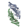 3rauS C: citing same article ( S: Starting model for refinement |
|---|---|
| Similar structure data |

-Links
 PDBj
PDBj


- Assembly
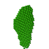
| Deposited unit | 
| ||||||||
|---|---|---|---|---|---|---|---|---|---|
| 1 | 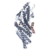
| ||||||||
| 2 | 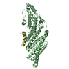
| ||||||||
| Unit cell |
|
-Components
| #1: Protein | Mass: 40584.816 Da / Num. of mol.: 2 / Fragment: UNP residues 1-361 / Mutation: N33A,Y34A Source method: isolated from a genetically manipulated source Source: (gene. exp.) Homo sapiens (human) / Gene: PTPN23, KIAA1471 / Production host:  Escherichia coli (E. coli) / References: UniProt: Q9H3S7, protein-tyrosine-phosphatase Escherichia coli (E. coli) / References: UniProt: Q9H3S7, protein-tyrosine-phosphatase#2: Protein/peptide | Mass: 2370.781 Da / Num. of mol.: 2 / Fragment: UNP residues 350-370 / Source method: obtained synthetically / Source: (synth.) Homo sapiens (human) / References: UniProt: O75886#3: Chemical | ChemComp-GOL / | Glycerol  Mass: 92.094 Da / Num. of mol.: 1 / Source method: obtained synthetically / Formula: C3H8O3 Mass: 92.094 Da / Num. of mol.: 1 / Source method: obtained synthetically / Formula: C3H8O3#4: Water | ChemComp-HOH / | Water Mass: 18.015 Da / Num. of mol.: 365 / Source method: isolated from a natural source / Formula: H2O Mass: 18.015 Da / Num. of mol.: 365 / Source method: isolated from a natural source / Formula: H2O |
|---|
-Experimental details
-Experiment
| Experiment | Method: X-RAY DIFFRACTION |
|---|
- Sample preparation
| Crystal | Density Matthews: 2.55 Å3/Da / Density % sol: 51.75 % |
|---|---|
| Crystal grow | Temperature: 293 K / Method: vapor diffusion, sitting drop / pH: 7 Details: 0.1 M Bis-Tris, 30%(w/v) polyethylene glycol 3350, 4%(v/v) acetonitrile |
-Data collection
| Diffraction | Mean temperature: 93 K |
|---|---|
| Diffraction source | Source: SYNCHROTRON / Site: PAL/PLS  / Beamline: 5C (4A) / Wavelength: 0.97949 Å / Beamline: 5C (4A) / Wavelength: 0.97949 Å |
| Detector | Type: ADSC QUANTUM 315r / Detector: CCD / Date: Nov 20, 2014 |
| Radiation | Protocol: SINGLE WAVELENGTH / Monochromatic (M) / Laue (L): M / Scattering type: x-ray |
| Radiation wavelength | Wavelength: 0.97949 Å / Relative weight: 1 |
| Reflection | Resolution: 2→50 Å / Num. obs: 56289 / % possible obs: 97.3 % / Redundancy: 4.8 % / Net I/σ(I): 30.4 |
| Reflection shell | Resolution: 2→2.03 Å / Mean I/σ(I) obs: 4.6 / % possible all: 95.5 |
- Processing
| Software |
| |||||||||||||||||||||||||||||||||||||||||||||||||||||||||||||||||||||||||||||||||||||||||||||||||||||||||
|---|---|---|---|---|---|---|---|---|---|---|---|---|---|---|---|---|---|---|---|---|---|---|---|---|---|---|---|---|---|---|---|---|---|---|---|---|---|---|---|---|---|---|---|---|---|---|---|---|---|---|---|---|---|---|---|---|---|---|---|---|---|---|---|---|---|---|---|---|---|---|---|---|---|---|---|---|---|---|---|---|---|---|---|---|---|---|---|---|---|---|---|---|---|---|---|---|---|---|---|---|---|---|---|---|---|---|
| Refinement | Method to determine structure: MOLECULAR REPLACEMENT Starting model: 3RAU Resolution: 2.001→29.089 Å / SU ML: 0.16 / Cross valid method: FREE R-VALUE / σ(F): 1.53 / Phase error: 23.89 / Stereochemistry target values: ML
| |||||||||||||||||||||||||||||||||||||||||||||||||||||||||||||||||||||||||||||||||||||||||||||||||||||||||
| Solvent computation | Shrinkage radii: 0.9 Å / VDW probe radii: 1.11 Å / Solvent model: FLAT BULK SOLVENT MODEL | |||||||||||||||||||||||||||||||||||||||||||||||||||||||||||||||||||||||||||||||||||||||||||||||||||||||||
| Refinement step | Cycle: LAST / Resolution: 2.001→29.089 Å
| |||||||||||||||||||||||||||||||||||||||||||||||||||||||||||||||||||||||||||||||||||||||||||||||||||||||||
| Refine LS restraints |
| |||||||||||||||||||||||||||||||||||||||||||||||||||||||||||||||||||||||||||||||||||||||||||||||||||||||||
| LS refinement shell |
|
