 Movie
Movie Controller

[English] 日本語
 Yorodumi
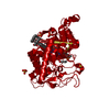
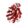
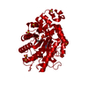
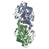
- PDB-5cpl: The crystal structure of Xenobiotic reductase A (XenA) from Pseud... -
+ Open data

- Basic information
| Entry | Database: PDB / ID: 5cpl | |||||||||
|---|---|---|---|---|---|---|---|---|---|---|
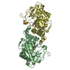
| Title | The crystal structure of Xenobiotic reductase A (XenA) from Pseudomonas putida in complex with a nicotinamide mimic (mNH2) | |||||||||
 Components Components | Xenobiotic reductase | |||||||||
 Keywords Keywords |  OXIDOREDUCTASE / Reductase / Nicotinamide mimic / biomimetic OXIDOREDUCTASE / Reductase / Nicotinamide mimic / biomimetic | |||||||||
| Function / homology |  Function and homology informationNADPH dehydrogenase activity / FMN binding / NADP binding / metal ion binding Function and homology informationNADPH dehydrogenase activity / FMN binding / NADP binding / metal ion bindingSimilarity search - Function | |||||||||
| Biological species |  Pseudomonas putida (bacteria) Pseudomonas putida (bacteria) | |||||||||
| Method | X-RAY DIFFRACTION / SYNCHROTRON / MOLECULAR REPLACEMENT / Resolution: 1.57 Å | |||||||||
 Authors Authors | Knaus, T. / Paul, C.E. / Levy, C.W. / Mutti, F.G. / Hollmann, F. / Scrutton, N.S. | |||||||||
| Funding support |  United Kingdom, 2items United Kingdom, 2items
| |||||||||
 Citation Citation | Journal: J.Am.Chem.Soc. / Year: 2016 Title: Better than Nature: Nicotinamide Biomimetics That Outperform Natural Coenzymes. Authors: Knaus, T. / Paul, C.E. / Levy, C.W. / de Vries, S. / Mutti, F.G. / Hollmann, F. / Scrutton, N.S. | |||||||||
| History |
|
- Structure visualization
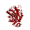
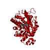
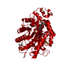
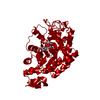
| Structure viewer | Molecule: MolmilJmol/JSmol |
|---|
- Downloads & links
-Download
| PDBx/mmCIF format | 5cpl.cif.gz | 290.5 KB | Display | PDBx/mmCIF format |
|---|---|---|---|---|
| PDB format | pdb5cpl.ent.gz | 236.1 KB | Display | PDB format |
| PDBx/mmJSON format | 5cpl.json.gz | Tree view | PDBx/mmJSON format | |
| Others |  Other downloads Other downloads |
-Validation report
| Arichive directory | https://data.pdbj.org/pub/pdb/validation_reports/cp/5cplftp://data.pdbj.org/pub/pdb/validation_reports/cp/5cpl | HTTPS FTP |
|---|
-Related structure data
| Related structure data |  5cpmC 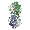 5cpnC  5cpoC  2h90S S: Starting model for refinement C: citing same article ( |
|---|---|
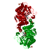
| Similar structure data |


-Links
 PDBj
PDBj- Assembly
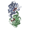
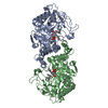
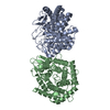
| Deposited unit | 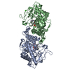
| ||||||||
|---|---|---|---|---|---|---|---|---|---|
| 1 |
| ||||||||
| Unit cell |
|
-Components
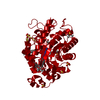
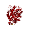
| #1: Protein | Mass: 40964.086 Da / Num. of mol.: 2 Source method: isolated from a genetically manipulated source Source: (gene. exp.) Pseudomonas putida (bacteria) / Gene: xenA / Plasmid: pET21A / Production host: Escherichia coli (E. coli) / References: UniProt: Q9R9V9#2: Chemical |   Mass: 216.279 Da / Num. of mol.: 2 / Source method: obtained synthetically / Formula: C13H16N2O Mass: 216.279 Da / Num. of mol.: 2 / Source method: obtained synthetically / Formula: C13H16N2O#3: Chemical | 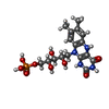  Mass: 458.360 Da / Num. of mol.: 2 / Source method: obtained synthetically / Formula: C17H23N4O9P Mass: 458.360 Da / Num. of mol.: 2 / Source method: obtained synthetically / Formula: C17H23N4O9P#4: Chemical | ChemComp-CA / |   Mass: 40.078 Da / Num. of mol.: 1 / Source method: obtained synthetically / Formula: Ca Mass: 40.078 Da / Num. of mol.: 1 / Source method: obtained synthetically / Formula: Ca#5: Water | ChemComp-HOH / | Water Mass: 18.015 Da / Num. of mol.: 797 / Source method: isolated from a natural source / Formula: H2O Mass: 18.015 Da / Num. of mol.: 797 / Source method: isolated from a natural source / Formula: H2O |
|---|
-Experimental details
-Experiment
| Experiment | Method: X-RAY DIFFRACTION |
|---|
- Sample preparation
| Crystal | Density Matthews: 2.26 Å3/Da / Density % sol: 45.69 % |
|---|---|
| Crystal grow | Temperature: 294 K / Method: vapor diffusion, sitting drop / pH: 7 Details: 0.2 M calcium acetate, 0.1 M sodium cacodylate buffer pH 6.5, 40% v/v PEG 300 (JCSG+ HT96 A10 Molecular Dimensions) Temp details: Incubator |
-Data collection
| Diffraction | Mean temperature: 100 K |
|---|---|
| Diffraction source | Source: SYNCHROTRON / Site: Diamond / Beamline: I03 / Wavelength: 0.9795 Å |
| Detector | Type: DECTRIS PILATUS 6M / Detector: PIXEL / Date: Jan 31, 2015 |
| Radiation | Protocol: SINGLE WAVELENGTH / Monochromatic (M) / Laue (L): M / Scattering type: x-ray |
| Radiation wavelength | Wavelength: 0.9795 Å / Relative weight: 1 |
| Reflection | Resolution: 1.57→47.02 Å / Num. all: 102314 / Num. obs: 102314 / % possible obs: 98 % / Redundancy: 6.7 % / Rmerge(I) obs: 0.0679 / Net I/σ(I): 17.41 |
| Reflection shell | Resolution: 1.57→1.626 Å / Redundancy: 6.7 % / Rmerge(I) obs: 0.669 / Mean I/σ(I) obs: 2.91 / % possible all: 96 |
- Processing
| Software |
| |||||||||||||||||||||||||||||||||||||||||||||||||||||||||||||||||||||||||||||||||||||||||||||||||||||||||||||||||||||||||||||||||||||||||||||||||||||||||||||||||||||||||||||||||||||||||||||||||||||||||||||||||||||||||
|---|---|---|---|---|---|---|---|---|---|---|---|---|---|---|---|---|---|---|---|---|---|---|---|---|---|---|---|---|---|---|---|---|---|---|---|---|---|---|---|---|---|---|---|---|---|---|---|---|---|---|---|---|---|---|---|---|---|---|---|---|---|---|---|---|---|---|---|---|---|---|---|---|---|---|---|---|---|---|---|---|---|---|---|---|---|---|---|---|---|---|---|---|---|---|---|---|---|---|---|---|---|---|---|---|---|---|---|---|---|---|---|---|---|---|---|---|---|---|---|---|---|---|---|---|---|---|---|---|---|---|---|---|---|---|---|---|---|---|---|---|---|---|---|---|---|---|---|---|---|---|---|---|---|---|---|---|---|---|---|---|---|---|---|---|---|---|---|---|---|---|---|---|---|---|---|---|---|---|---|---|---|---|---|---|---|---|---|---|---|---|---|---|---|---|---|---|---|---|---|---|---|---|---|---|---|---|---|---|---|---|---|---|---|---|---|---|---|---|
| Refinement | Method to determine structure: MOLECULAR REPLACEMENT Starting model: 2H90 Resolution: 1.57→47.02 Å / SU ML: 0.12 / Cross valid method: FREE R-VALUE / σ(F): 0 / Phase error: 14.88 / Stereochemistry target values: ML
| |||||||||||||||||||||||||||||||||||||||||||||||||||||||||||||||||||||||||||||||||||||||||||||||||||||||||||||||||||||||||||||||||||||||||||||||||||||||||||||||||||||||||||||||||||||||||||||||||||||||||||||||||||||||||
| Solvent computation | Shrinkage radii: 0.9 Å / VDW probe radii: 1.11 Å / Solvent model: FLAT BULK SOLVENT MODEL | |||||||||||||||||||||||||||||||||||||||||||||||||||||||||||||||||||||||||||||||||||||||||||||||||||||||||||||||||||||||||||||||||||||||||||||||||||||||||||||||||||||||||||||||||||||||||||||||||||||||||||||||||||||||||
| Refinement step | Cycle: LAST / Resolution: 1.57→47.02 Å
| |||||||||||||||||||||||||||||||||||||||||||||||||||||||||||||||||||||||||||||||||||||||||||||||||||||||||||||||||||||||||||||||||||||||||||||||||||||||||||||||||||||||||||||||||||||||||||||||||||||||||||||||||||||||||
| Refine LS restraints |
| |||||||||||||||||||||||||||||||||||||||||||||||||||||||||||||||||||||||||||||||||||||||||||||||||||||||||||||||||||||||||||||||||||||||||||||||||||||||||||||||||||||||||||||||||||||||||||||||||||||||||||||||||||||||||
| LS refinement shell |
|
