 Movie
Movie Controller

[English] 日本語
 Yorodumi
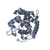
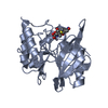
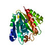
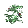
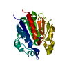
- PDB-5cmt: Fic protein from Neisseria meningitidis (NmFic) mutant E156R Y183... -
+ Open data

- Basic information
| Entry | Database: PDB / ID: 5cmt | ||||||
|---|---|---|---|---|---|---|---|
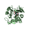
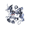
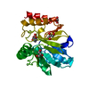
| Title | Fic protein from Neisseria meningitidis (NmFic) mutant E156R Y183F in dimeric form | ||||||
 Components Components | Adenosine monophosphate-protein transferase NmFic | ||||||
 Keywords Keywords |  TRANSFERASE / Fic protein / AMP-transferase / dimer TRANSFERASE / Fic protein / AMP-transferase / dimer | ||||||
| Function / homology |  Function and homology information Function and homology informationAMPylase activity / protein adenylyltransferase / protein adenylylation / regulation of cell division / protein homodimerization activity / ATP bindingSimilarity search - Function | ||||||
| Biological species |  Neisseria meningitidis serogroup B (bacteria) Neisseria meningitidis serogroup B (bacteria) | ||||||
| Method | X-RAY DIFFRACTION / SYNCHROTRON / MOLECULAR REPLACEMENT / molecular replacement / Resolution: 0.99 Å | ||||||
 Authors Authors | Stanger, F.V. / Schirmer, T. | ||||||
 Citation Citation | Journal: Proc.Natl.Acad.Sci.USA / Year: 2016 Title: Intrinsic regulation of FIC-domain AMP-transferases by oligomerization and automodification. Authors: Stanger, F.V. / Burmann, B.M. / Harms, A. / Aragao, H. / Mazur, A. / Sharpe, T. / Dehio, C. / Hiller, S. / Schirmer, T. | ||||||
| History |
|
- Structure visualization
| Structure viewer | Molecule: MolmilJmol/JSmol |
|---|
- Downloads & links
-Download
| PDBx/mmCIF format | 5cmt.cif.gz | 106 KB | Display | PDBx/mmCIF format |
|---|---|---|---|---|
| PDB format | pdb5cmt.ent.gz | 81.2 KB | Display | PDB format |
| PDBx/mmJSON format | 5cmt.json.gz | Tree view | PDBx/mmJSON format | |
| Others |  Other downloads Other downloads |
-Validation report
| Arichive directory | https://data.pdbj.org/pub/pdb/validation_reports/cm/5cmtftp://data.pdbj.org/pub/pdb/validation_reports/cm/5cmt | HTTPS FTP |
|---|
-Related structure data
| Related structure data |  5cglC 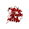 5cklSC S: Starting model for refinement C: citing same article ( |
|---|---|
| Similar structure data |


-Links
 PDBj
PDBj- Assembly
| Deposited unit | 
| ||||||||
|---|---|---|---|---|---|---|---|---|---|
| 1 |
| ||||||||
| Unit cell |
| ||||||||
| Components on special symmetry positions |
|
-Components
| #1: Protein | Mass: 22135.230 Da / Num. of mol.: 1 / Fragment: UNP residues 21-191 / Mutation: E156R Y183F Source method: isolated from a genetically manipulated source Source: (gene. exp.) Neisseria meningitidis serogroup B (bacteria)Gene: NMB0255 / Plasmid: pRSFDuet1 / Production host: Escherichia coli (E. coli) / Strain (production host): BL21(DE3)References: UniProt: Q7DDR9, Transferases; Transferring phosphorus-containing groups; Nucleotidyltransferases | ||||
|---|---|---|---|---|---|
| #2: Chemical | Glycerol  Mass: 92.094 Da / Num. of mol.: 2 / Source method: obtained synthetically / Formula: C3H8O3 Mass: 92.094 Da / Num. of mol.: 2 / Source method: obtained synthetically / Formula: C3H8O3#3: Chemical | Chloride  Mass: 35.453 Da / Num. of mol.: 3 / Source method: obtained synthetically / Formula: Cl Mass: 35.453 Da / Num. of mol.: 3 / Source method: obtained synthetically / Formula: Cl#4: Water | ChemComp-HOH / | Water Mass: 18.015 Da / Num. of mol.: 248 / Source method: isolated from a natural source / Formula: H2O Mass: 18.015 Da / Num. of mol.: 248 / Source method: isolated from a natural source / Formula: H2O |
-Experimental details
-Experiment
| Experiment | Method: X-RAY DIFFRACTION / Number of used crystals: 1 |
|---|
- Sample preparation
| Crystal | Density Matthews: 2.74 Å3/Da / Density % sol: 54.79 % |
|---|---|
| Crystal grow | Temperature: 277.15 K / Method: batch mode / pH: 7.8 / Details: 10 mM Tris pH 7.8, 100 mM NaCl |
-Data collection
| Diffraction | Mean temperature: 100 K | |||||||||||||||||||||||||||
|---|---|---|---|---|---|---|---|---|---|---|---|---|---|---|---|---|---|---|---|---|---|---|---|---|---|---|---|---|
| Diffraction source | Source: SYNCHROTRON / Site: SLS  / Beamline: X06DA / Wavelength: 0.8 Å / Beamline: X06DA / Wavelength: 0.8 Å | |||||||||||||||||||||||||||
| Detector | Type: DECTRIS PILATUS 2M / Detector: PIXEL / Date: Feb 11, 2013 | |||||||||||||||||||||||||||
| Radiation | Protocol: SINGLE WAVELENGTH / Monochromatic (M) / Laue (L): M / Scattering type: x-ray | |||||||||||||||||||||||||||
| Radiation wavelength | Wavelength: 0.8 Å / Relative weight: 1 | |||||||||||||||||||||||||||
| Reflection | Resolution: 0.99→47.22 Å / Num. obs: 126436 / % possible obs: 96.8 % / Redundancy: 6.4 % / CC1/2: 0.999 / Rmerge(I) obs: 0.04 / Rpim(I) all: 0.017 / Net I/σ(I): 23.4 / Num. measured all: 805863 / Scaling rejects: 135 | |||||||||||||||||||||||||||
| Reflection shell | Diffraction-ID: 1 / Rejects: 0
|
-Phasing
| Phasing | Method: molecular replacement | |||||||||
|---|---|---|---|---|---|---|---|---|---|---|
| Phasing MR | Model details: Phaser MODE: MR_AUTO
|
- Processing
| Software |
| ||||||||||||||||||||||||||||||||||||||||||||||||||||||||||||||||||||||||||||||||||||||||||
|---|---|---|---|---|---|---|---|---|---|---|---|---|---|---|---|---|---|---|---|---|---|---|---|---|---|---|---|---|---|---|---|---|---|---|---|---|---|---|---|---|---|---|---|---|---|---|---|---|---|---|---|---|---|---|---|---|---|---|---|---|---|---|---|---|---|---|---|---|---|---|---|---|---|---|---|---|---|---|---|---|---|---|---|---|---|---|---|---|---|---|---|
| Refinement | Method to determine structure: MOLECULAR REPLACEMENT Starting model: 5CKL Resolution: 0.99→47.22 Å / Cor.coef. Fo:Fc: 0.977 / Cor.coef. Fo:Fc free: 0.971 / WRfactor Rfree: 0.1479 / WRfactor Rwork: 0.1273 / FOM work R set: 0.9139 / SU B: 0.595 / SU ML: 0.014 / SU R Cruickshank DPI: 0.0186 / SU Rfree: 0.0199 / Cross valid method: THROUGHOUT / σ(F): 0 / ESU R: 0.019 / ESU R Free: 0.02 / Stereochemistry target values: MAXIMUM LIKELIHOOD Details: HYDROGENS HAVE BEEN ADDED IN THE RIDING POSITIONS U VALUES : REFINED INDIVIDUALLY
| ||||||||||||||||||||||||||||||||||||||||||||||||||||||||||||||||||||||||||||||||||||||||||
| Solvent computation | Ion probe radii: 0.8 Å / Shrinkage radii: 0.8 Å / VDW probe radii: 1.2 Å / Solvent model: MASK | ||||||||||||||||||||||||||||||||||||||||||||||||||||||||||||||||||||||||||||||||||||||||||
| Displacement parameters | Biso max: 86.39 Å2 / Biso mean: 11.055 Å2 / Biso min: 4.19 Å2
| ||||||||||||||||||||||||||||||||||||||||||||||||||||||||||||||||||||||||||||||||||||||||||
| Refinement step | Cycle: final / Resolution: 0.99→47.22 Å
| ||||||||||||||||||||||||||||||||||||||||||||||||||||||||||||||||||||||||||||||||||||||||||
| Refine LS restraints |
| ||||||||||||||||||||||||||||||||||||||||||||||||||||||||||||||||||||||||||||||||||||||||||
| LS refinement shell | Resolution: 0.99→1.016 Å / Total num. of bins used: 20
|
