 Movie
Movie Controller


+ Open data

- Basic information
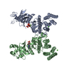
| Entry | Database: PDB / ID: 5clr | ||||||
|---|---|---|---|---|---|---|---|
| Title | Crystal structure of LegK4_APO Kinase | ||||||
 Components Components | LegK4 | ||||||
 Keywords Keywords |  TRANSFERASE / Legionella / Bacterial effectors / serine/threonine kinase / type IV secretion system TRANSFERASE / Legionella / Bacterial effectors / serine/threonine kinase / type IV secretion system | ||||||
| Function / homology |  Function and homology informationnon-specific serine/threonine protein kinase / protein phosphorylation / protein serine/threonine kinase activity / ATP binding Function and homology informationnon-specific serine/threonine protein kinase / protein phosphorylation / protein serine/threonine kinase activity / ATP bindingSimilarity search - Function | ||||||
| Biological species |   Legionella pneumophila (bacteria) Legionella pneumophila (bacteria) | ||||||
| Method | X-RAY DIFFRACTION / SYNCHROTRON / SAD / Resolution: 3.706 Å | ||||||
 Authors Authors | Flayhan, A. / Terradot, L. | ||||||
 Citation Citation | Journal: Sci Rep / Year: 2015 Title: The structure of Legionella pneumophila LegK4 type four secretion system (T4SS) effector reveals a novel dimeric eukaryotic-like kinase. Authors: Flayhan, A. / Berge, C. / Bailo, N. / Doublet, P. / Bayliss, R. / Terradot, L. | ||||||
| History |
|
- Structure visualization
| Structure viewer | Molecule: MolmilJmol/JSmol |
|---|
- Downloads & links
-Download
| PDBx/mmCIF format | 5clr.cif.gz | 291.5 KB | Display | PDBx/mmCIF format |
|---|---|---|---|---|
| PDB format | pdb5clr.ent.gz | 248.1 KB | Display | PDB format |
| PDBx/mmJSON format | 5clr.json.gz | Tree view | PDBx/mmJSON format | |
| Others |  Other downloads Other downloads |
-Validation report
| Arichive directory | https://data.pdbj.org/pub/pdb/validation_reports/cl/5clrftp://data.pdbj.org/pub/pdb/validation_reports/cl/5clr | HTTPS FTP |
|---|
-Related structure data


-Links
 PDBj
PDBj
- Assembly
| Deposited unit | 
| ||||||||
|---|---|---|---|---|---|---|---|---|---|
| 1 |
| ||||||||
| Unit cell |
|
-Components
| #1: Protein | Mass: 51578.902 Da / Num. of mol.: 2 Source method: isolated from a genetically manipulated source Source: (gene. exp.) Legionella pneumophila (strain Lens) (bacteria)Gene: lpl0262 / Production host: Escherichia coli BL21(DE3) (bacteria) / References: UniProt: Q5WZW9 |
|---|
-Experimental details
-Experiment
| Experiment | Method: X-RAY DIFFRACTION |
|---|
- Sample preparation
| Crystal | Density Matthews: 3.07 Å3/Da / Density % sol: 59.97 % |
|---|---|
| Crystal grow | Temperature: 293 K / Method: vapor diffusion, hanging drop / pH: 7.5 Details: 12% (w/v) PEG 4000, 20% glycerol, 0.1 M MOPS/HEPES pH 7.5, 5 mM Hexaammine cobalt (III) chloride. |
-Data collection
| Diffraction | Mean temperature: 173 K |
|---|---|
| Diffraction source | Source: SYNCHROTRON / Site: SOLEIL  / Beamline: PROXIMA 2 / Wavelength: 0.979 Å / Beamline: PROXIMA 2 / Wavelength: 0.979 Å |
| Detector | Type: ADSC QUANTUM 315r / Detector: CCD / Date: Nov 16, 2013 |
| Radiation | Monochromator: si [111] crystal / Protocol: SINGLE WAVELENGTH / Monochromatic (M) / Laue (L): M / Scattering type: x-ray |
| Radiation wavelength | Wavelength: 0.979 Å / Relative weight: 1 |
| Reflection | Resolution: 3.7→44.693 Å / Num. obs: 13215 / % possible obs: 100 % / Redundancy: 11 % / Rmerge(I) obs: 0.239 / Net I/σ(I): 13.79 |
| Reflection shell | Resolution: 3.706→3.838 Å / Redundancy: 2.47 % / Rmerge(I) obs: 1.419 / % possible all: 100 |
- Processing
| Software |
| ||||||||||||||||||||||||||||||||||||||||||
|---|---|---|---|---|---|---|---|---|---|---|---|---|---|---|---|---|---|---|---|---|---|---|---|---|---|---|---|---|---|---|---|---|---|---|---|---|---|---|---|---|---|---|---|
| Refinement | Method to determine structure: SAD / Resolution: 3.706→44.693 Å / SU ML: 0.59 / Cross valid method: FREE R-VALUE / σ(F): 1.35 / Phase error: 34.22 / Stereochemistry target values: ML
| ||||||||||||||||||||||||||||||||||||||||||
| Solvent computation | Shrinkage radii: 0.9 Å / VDW probe radii: 1.11 Å / Solvent model: FLAT BULK SOLVENT MODEL | ||||||||||||||||||||||||||||||||||||||||||
| Refinement step | Cycle: LAST / Resolution: 3.706→44.693 Å
| ||||||||||||||||||||||||||||||||||||||||||
| Refine LS restraints |
| ||||||||||||||||||||||||||||||||||||||||||
| LS refinement shell |
|
