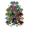
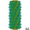
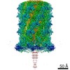
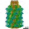
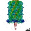
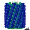
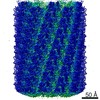
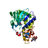
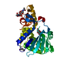
Journal: Nature / Year: 2020 Title: Action of a minimal contractile bactericidal nanomachine. Authors: Peng Ge / Dean Scholl / Nikolai S Prokhorov / Jaycob Avaylon / Mikhail M Shneider / Christopher Browning / Sergey A Buth / Michel Plattner / Urmi Chakraborty / Ke Ding / Petr G Leiman / Jeff ...Authors: Peng Ge / Dean Scholl / Nikolai S Prokhorov / Jaycob Avaylon / Mikhail M Shneider / Christopher Browning / Sergey A Buth / Michel Plattner / Urmi Chakraborty / Ke Ding / Petr G Leiman / Jeff F Miller / Z Hong Zhou / Abstract: R-type bacteriocins are minimal contractile nanomachines that hold promise as precision antibiotics. Each bactericidal complex uses a collar to bridge a hollow tube with a contractile sheath loaded ...R-type bacteriocins are minimal contractile nanomachines that hold promise as precision antibiotics. Each bactericidal complex uses a collar to bridge a hollow tube with a contractile sheath loaded in a metastable state by a baseplate scaffold. Fine-tuning of such nucleic acid-free protein machines for precision medicine calls for an atomic description of the entire complex and contraction mechanism, which is not available from baseplate structures of the (DNA-containing) T4 bacteriophage. Here we report the atomic model of the complete R2 pyocin in its pre-contraction and post-contraction states, each containing 384 subunits of 11 unique atomic models of 10 gene products. Comparison of these structures suggests the following sequence of events during pyocin contraction: tail fibres trigger lateral dissociation of baseplate triplexes; the dissociation then initiates a cascade of events leading to sheath contraction; and this contraction converts chemical energy into mechanical force to drive the iron-tipped tube across the bacterial cell surface, killing the bacterium.
In the structure databanks used in Yorodumi, some data are registered as the other names, "COVID-19 virus" and "2019-nCoV". Here are the details of the virus and the list of structure data.
Jan 31, 2019. EMDB accession codes are about to change! (news from PDBe EMDB page)
EMDB accession codes are about to change! (news from PDBe EMDB page)
The allocation of 4 digits for EMDB accession codes will soon come to an end. Whilst these codes will remain in use, new EMDB accession codes will include an additional digit and will expand incrementally as the available range of codes is exhausted. The current 4-digit format prefixed with “EMD-” (i.e. EMD-XXXX) will advance to a 5-digit format (i.e. EMD-XXXXX), and so on. It is currently estimated that the 4-digit codes will be depleted around Spring 2019, at which point the 5-digit format will come into force.
The EM Navigator/Yorodumi systems omit the EMD- prefix.
Related info.:Q: What is EMD? / ID/Accession-code notation in Yorodumi/EM Navigator
Yorodumi is a browser for structure data from EMDB, PDB, SASBDB, etc.
This page is also the successor to EM Navigator detail page, and also detail information page/front-end page for Omokage search.
The word "yorodu" (or yorozu) is an old Japanese word meaning "ten thousand". "mi" (miru) is to see.
Related info.:EMDB / PDB / SASBDB / Comparison of 3 databanks / Yorodumi Search / Aug 31, 2016. New EM Navigator & Yorodumi / Yorodumi Papers / Jmol/JSmol / Function and homology information / Changes in new EM Navigator and Yorodumi
 Movie
Movie Controller


 Open data
 Basic information
 Components
 Keywords
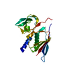
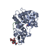
 STRUCTURAL PROTEIN / gpJ / gp6
 Function and homology information
 Authors
 Switzerland, 1items
  Citation


 Structure visualization
 Downloads & links
 Other downloads


 PDBj
PDBj Assembly
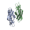
 Mass: 18.015 Da / Num. of mol.: 58 / Source method: isolated from a natural source / Formula: H2O
 Sample preparation
 Processing