 Movie
Movie Controller

[English] 日本語
 Yorodumi
- PDB-5c7k: Crystal structure BG505 SOSIP gp140 HIV-1 Env trimer bound to bro... -
+ Open data

- Basic information
| Entry | Database: PDB / ID: 5c7k | |||||||||
|---|---|---|---|---|---|---|---|---|---|---|
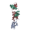
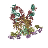
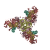
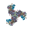
| Title | Crystal structure BG505 SOSIP gp140 HIV-1 Env trimer bound to broadly neutralizing antibodies PGT128 and 8ANC195 | |||||||||
 Components Components |
| |||||||||
 Keywords Keywords |  IMMUNE SYSTEM / HIV-1 / SOSIP / PGT128 / 8ANC195 IMMUNE SYSTEM / HIV-1 / SOSIP / PGT128 / 8ANC195 | |||||||||
| Function / homology |  Function and homology information Function and homology informationIgD immunoglobulin complex / IgM immunoglobulin complex / IgA immunoglobulin complex / IgE immunoglobulin complex / CD22 mediated BCR regulation / complement-dependent cytotoxicity / Fc epsilon receptor (FCERI) signaling / antibody-dependent cellular cytotoxicity / Fc-gamma receptor I complex binding / Classical antibody-mediated complement activation ...IgD immunoglobulin complex / IgM immunoglobulin complex / IgA immunoglobulin complex / IgE immunoglobulin complex / CD22 mediated BCR regulation / complement-dependent cytotoxicity / Fc epsilon receptor (FCERI) signaling / antibody-dependent cellular cytotoxicity / Fc-gamma receptor I complex binding / Classical antibody-mediated complement activation / IgG immunoglobulin complex / Initial triggering of complement / immunoglobulin complex / immunoglobulin complex, circulating / immunoglobulin receptor binding / immunoglobulin mediated immune response / FCGR activation / Role of phospholipids in phagocytosis / Role of LAT2/NTAL/LAB on calcium mobilization / Scavenging of heme from plasma / positive regulation of plasma membrane raft polarization / positive regulation of receptor clustering / complement activation, classical pathway / positive regulation of establishment of T cell polarity / antigen binding / FCERI mediated Ca+2 mobilization / FCGR3A-mediated IL10 synthesis / host cell endosome membrane / Antigen activates B Cell Receptor (BCR) leading to generation of second messengers / Regulation of Complement cascade / Cell surface interactions at the vascular wall / FCERI mediated MAPK activation / FCGR3A-mediated phagocytosis / B cell receptor signaling pathway / Regulation of actin dynamics for phagocytic cup formation / FCERI mediated NF-kB activation / Immunoregulatory interactions between a Lymphoid and a non-Lymphoid cell / antibacterial humoral response / clathrin-dependent endocytosis of virus by host cell / Interleukin-4 and Interleukin-13 signaling / Potential therapeutics for SARS / adaptive immune response / blood microparticle / viral protein processing / virus-mediated perturbation of host defense response / immune response / fusion of virus membrane with host plasma membrane / fusion of virus membrane with host endosome membrane / viral envelope / virion attachment to host cell / host cell plasma membrane / virion membrane / structural molecule activity / extracellular space / extracellular exosome / extracellular region / identical protein binding / plasma membraneSimilarity search - Function | |||||||||
| Biological species |  Homo sapiens (human) Homo sapiens (human)  Human immunodeficiency virus 1 Human immunodeficiency virus 1 | |||||||||
| Method | X-RAY DIFFRACTION / SYNCHROTRON / MOLECULAR REPLACEMENT / Resolution: 4.6019 Å | |||||||||
 Authors Authors | Kong, L. / Stanfield, R.L. / Wilson, I.A. | |||||||||
 Citation Citation | Journal: Acta Crystallogr.,Sect.D / Year: 2015 Title: Complete epitopes for vaccine design derived from a crystal structure of the broadly neutralizing antibodies PGT128 and 8ANC195 in complex with an HIV-1 Env trimer. Authors: Kong, L. / Torrents de la Pena, A. / Deller, M.C. / Garces, F. / Sliepen, K. / Hua, Y. / Stanfield, R.L. / Sanders, R.W. / Wilson, I.A. | |||||||||
| History |
|
- Structure visualization
| Structure viewer | Molecule: MolmilJmol/JSmol |
|---|
- Downloads & links
-Download
| PDBx/mmCIF format | 5c7k.cif.gz | 288.1 KB | Display | PDBx/mmCIF format |
|---|---|---|---|---|
| PDB format | pdb5c7k.ent.gz | 232.1 KB | Display | PDB format |
| PDBx/mmJSON format | 5c7k.json.gz | Tree view | PDBx/mmJSON format | |
| Others |  Other downloads Other downloads |
-Validation report
| Arichive directory | https://data.pdbj.org/pub/pdb/validation_reports/c7/5c7kftp://data.pdbj.org/pub/pdb/validation_reports/c7/5c7k | HTTPS FTP |
|---|
-Related structure data


-Links
 PDBj
PDBj


- Assembly
| Deposited unit | 
| ||||||||
|---|---|---|---|---|---|---|---|---|---|
| 1 | 
| ||||||||
| Unit cell |
| ||||||||
| Details | The biological unit consists of three gp140 subunits (each consisting of one gp41 and one gp120 chain) each bound to antibodies PGT128 and 8ANC195. This is evidenced by gel filtration chromatography. |
-Components
-Envelope glycoprotein ... , 2 types, 2 molecules CD
| #3: Protein | Mass: 54596.672 Da / Num. of mol.: 1 / Fragment: UNP Residues 30-507 Source method: isolated from a genetically manipulated source Source: (gene. exp.) Human immunodeficiency virus 1 / Gene: env / Production host: Homo sapiens (human) / References: UniProt: Q2N0S6 |
|---|---|
| #4: Protein | Mass: 17146.482 Da / Num. of mol.: 1 Source method: isolated from a genetically manipulated source Source: (gene. exp.) Human immunodeficiency virus 1 / Gene: env / Cell line (production host): HEK 293 / Production host: Homo sapiens (human) / References: UniProt: Q2N0S6 |
-Antibody , 4 types, 4 molecules ABEF
| #1: Antibody | Mass: 25580.701 Da / Num. of mol.: 1 Source method: isolated from a genetically manipulated source Source: (gene. exp.) Homo sapiens (human) / Cell line (production host): HEK 293 / Production host: Homo sapiens (human) / References: UniProt: S6B291 |
|---|---|
| #2: Antibody | Mass: 22223.588 Da / Num. of mol.: 1 Source method: isolated from a genetically manipulated source Source: (gene. exp.) Homo sapiens (human) / Gene: IgLC-rG / Production host: Homo sapiens (human) / References: UniProt: C6KXN3 |
| #5: Antibody | Mass: 25268.375 Da / Num. of mol.: 1 Source method: isolated from a genetically manipulated source Source: (gene. exp.) Homo sapiens (human) / Gene: IGHG1 / Cell line (production host): HEK 293 / Production host: Homo sapiens (human) / References: UniProt: P01857 |
| #6: Antibody | Mass: 23401.984 Da / Num. of mol.: 1 Source method: isolated from a genetically manipulated source Source: (gene. exp.) Homo sapiens (human) / Gene: IGKC / Cell line (production host): HEK 293 / Production host: Homo sapiens (human) / References: UniProt: P01834 |
-Sugars , 9 types, 20 molecules 
| #7: Polysaccharide | 2-acetamido-2-deoxy-beta-D-glucopyranose-(1-4)-2-acetamido-2-deoxy-beta-D-glucopyranose / Mass: 424.401 Da / Num. of mol.: 4Source method: isolated from a genetically manipulated source #8: Polysaccharide | / Mass: 1559.386 Da / Num. of mol.: 2Source method: isolated from a genetically manipulated source #9: Polysaccharide | alpha-D-mannopyranose-(1-2)-alpha-D-mannopyranose-(1-2)-alpha-D-mannopyranose-(1-3)-[alpha-D- ...alpha-D-mannopyranose-(1-2)-alpha-D-mannopyranose-(1-2)-alpha-D-mannopyranose-(1-3)-[alpha-D-mannopyranose-(1-2)-alpha-D-mannopyranose-(1-6)-[alpha-D-mannopyranose-(1-3)]alpha-D-mannopyranose-(1-6)]beta-D-mannopyranose-(1-4)-2-acetamido-2-deoxy-beta-D-glucopyranose-(1-4)-2-acetamido-2-deoxy-beta-D-glucopyranose | / Mass: 1721.527 Da / Num. of mol.: 1Source method: isolated from a genetically manipulated source #10: Polysaccharide | alpha-D-mannopyranose-(1-2)-alpha-D-mannopyranose-(1-2)-alpha-D-mannopyranose-(1-3)-[alpha-D- ...alpha-D-mannopyranose-(1-2)-alpha-D-mannopyranose-(1-2)-alpha-D-mannopyranose-(1-3)-[alpha-D-mannopyranose-(1-6)]beta-D-mannopyranose-(1-4)-2-acetamido-2-deoxy-beta-D-glucopyranose-(1-4)-2-acetamido-2-deoxy-beta-D-glucopyranose | / Mass: 1235.105 Da / Num. of mol.: 1Source method: isolated from a genetically manipulated source #11: Polysaccharide | alpha-D-mannopyranose-(1-2)-alpha-D-mannopyranose-(1-3)-beta-D-mannopyranose-(1-4)-2-acetamido-2- ...alpha-D-mannopyranose-(1-2)-alpha-D-mannopyranose-(1-3)-beta-D-mannopyranose-(1-4)-2-acetamido-2-deoxy-beta-D-glucopyranose-(1-4)-2-acetamido-2-deoxy-beta-D-glucopyranose | / Mass: 910.823 Da / Num. of mol.: 1Source method: isolated from a genetically manipulated source #12: Polysaccharide | / Mass: 586.542 Da / Num. of mol.: 3Source method: isolated from a genetically manipulated source #13: Polysaccharide | alpha-D-mannopyranose-(1-2)-alpha-D-mannopyranose-(1-3)-[alpha-D-mannopyranose-(1-6)]alpha-D- ...alpha-D-mannopyranose-(1-2)-alpha-D-mannopyranose-(1-3)-[alpha-D-mannopyranose-(1-6)]alpha-D-mannopyranose-(1-6)-[alpha-D-mannopyranose-(1-3)]beta-D-mannopyranose-(1-4)-2-acetamido-2-deoxy-beta-D-glucopyranose-(1-4)-2-acetamido-2-deoxy-beta-D-glucopyranose | / Mass: 1397.245 Da / Num. of mol.: 1Source method: isolated from a genetically manipulated source #14: Polysaccharide | alpha-D-mannopyranose-(1-2)-alpha-D-mannopyranose-(1-2)-alpha-D-mannopyranose-(1-3)-[alpha-D- ...alpha-D-mannopyranose-(1-2)-alpha-D-mannopyranose-(1-2)-alpha-D-mannopyranose-(1-3)-[alpha-D-mannopyranose-(1-2)-alpha-D-mannopyranose-(1-3)-[alpha-D-mannopyranose-(1-2)-alpha-D-mannopyranose-(1-6)]alpha-D-mannopyranose-(1-6)]beta-D-mannopyranose-(1-4)-2-acetamido-2-deoxy-beta-D-glucopyranose-(1-4)-2-acetamido-2-deoxy-beta-D-glucopyranose | / Mass: 1883.668 Da / Num. of mol.: 1Source method: isolated from a genetically manipulated source #15: Sugar | ChemComp-NAG / N-Acetylglucosamine Type: D-saccharide, beta linking / Mass: 221.208 Da / Num. of mol.: 6 Type: D-saccharide, beta linking / Mass: 221.208 Da / Num. of mol.: 6Source method: isolated from a genetically manipulated source Formula: C8H15NO6 |
|---|
-Non-polymers , 1 types, 1 molecules 
| #16: Chemical | ChemComp-SO4 / Sulfate Mass: 96.063 Da / Num. of mol.: 1 / Source method: obtained synthetically / Formula: SO4 Mass: 96.063 Da / Num. of mol.: 1 / Source method: obtained synthetically / Formula: SO4 |
|---|
-Experimental details
-Experiment
| Experiment | Method: X-RAY DIFFRACTION |
|---|
- Sample preparation
| Crystal | Density Matthews: 4.42 Å3/Da / Density % sol: 72.18 % |
|---|---|
| Crystal grow | Temperature: 280 K / Method: vapor diffusion Details: 0.05 M lithium sulfate, 0.05 M sodium sulfate, 26% (w/v) PEG 400 and 0.1 M Tris-HCl pH 8.3 |
-Data collection
| Diffraction | Mean temperature: 100 K |
|---|---|
| Diffraction source | Source: SYNCHROTRON / Site: APS  / Beamline: 23-ID-B / Wavelength: 1.0332 Å / Beamline: 23-ID-B / Wavelength: 1.0332 Å |
| Detector | Type: MARMOSAIC 300 mm CCD / Detector: CCD / Date: Dec 11, 2014 |
| Radiation | Protocol: SINGLE WAVELENGTH / Monochromatic (M) / Laue (L): M / Scattering type: x-ray |
| Radiation wavelength | Wavelength: 1.0332 Å / Relative weight: 1 |
| Reflection | Resolution: 4.6→50 Å / Num. obs: 16539 / % possible obs: 99.8 % / Redundancy: 4.5 % / Rsym value: 0.21 / Net I/σ(I): 7.8 |
| Reflection shell | Resolution: 4.6→4.76 Å / Redundancy: 4.6 % / Rmerge(I) obs: 0.21 / Mean I/σ(I) obs: 1.7 / % possible all: 100 |
- Processing
| Software |
| |||||||||||||||||||||||||||||||||||||||||||||||||||||||||||||||||||||||||||||||||||||||||||
|---|---|---|---|---|---|---|---|---|---|---|---|---|---|---|---|---|---|---|---|---|---|---|---|---|---|---|---|---|---|---|---|---|---|---|---|---|---|---|---|---|---|---|---|---|---|---|---|---|---|---|---|---|---|---|---|---|---|---|---|---|---|---|---|---|---|---|---|---|---|---|---|---|---|---|---|---|---|---|---|---|---|---|---|---|---|---|---|---|---|---|---|---|
| Refinement | Method to determine structure: MOLECULAR REPLACEMENT Starting model: 4TVP, 3TYG, 4P9H Resolution: 4.6019→47.68 Å / SU ML: 0.62 / Cross valid method: FREE R-VALUE / σ(F): 1.34 / Phase error: 31.84 / Stereochemistry target values: ML
| |||||||||||||||||||||||||||||||||||||||||||||||||||||||||||||||||||||||||||||||||||||||||||
| Solvent computation | Shrinkage radii: 0.9 Å / VDW probe radii: 1.11 Å / Solvent model: FLAT BULK SOLVENT MODEL | |||||||||||||||||||||||||||||||||||||||||||||||||||||||||||||||||||||||||||||||||||||||||||
| Refinement step | Cycle: LAST / Resolution: 4.6019→47.68 Å
| |||||||||||||||||||||||||||||||||||||||||||||||||||||||||||||||||||||||||||||||||||||||||||
| Refine LS restraints |
| |||||||||||||||||||||||||||||||||||||||||||||||||||||||||||||||||||||||||||||||||||||||||||
| LS refinement shell |
|
