 Movie
Movie Controller

[English] 日本語
 Yorodumi
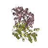
- PDB-5c5e: Structure of KaiA dimer in complex with C-terminal KaiC peptide a... -
+ Open data

- Basic information
| Entry | Database: PDB / ID: 5c5e | ||||||
|---|---|---|---|---|---|---|---|
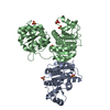
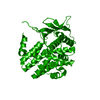
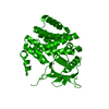
| Title | Structure of KaiA dimer in complex with C-terminal KaiC peptide at 2.8 A resolution | ||||||
 Components Components |
| ||||||
 Keywords Keywords |  TRANSCRIPTION / Clock protein KaiA-KaiC complex TRANSCRIPTION / Clock protein KaiA-KaiC complex | ||||||
| Function / homology |  Function and homology information Function and homology informationdetection of redox state / negative regulation of protein dephosphorylation / regulation of phosphorelay signal transduction system / negative regulation of circadian rhythm / entrainment of circadian clock / positive regulation of circadian rhythm / protein serine/threonine/tyrosine kinase activity / Hydrolases; Acting on acid anhydrides; Acting on acid anhydrides to facilitate cellular and subcellular movement / circadian rhythm / non-specific serine/threonine protein kinase ...detection of redox state / negative regulation of protein dephosphorylation / regulation of phosphorelay signal transduction system / negative regulation of circadian rhythm / entrainment of circadian clock / positive regulation of circadian rhythm / protein serine/threonine/tyrosine kinase activity / Hydrolases; Acting on acid anhydrides; Acting on acid anhydrides to facilitate cellular and subcellular movement / circadian rhythm / non-specific serine/threonine protein kinase / phosphorylation / protein serine kinase activity / protein serine/threonine kinase activity / regulation of DNA-templated transcription / magnesium ion binding / ATP hydrolysis activity / DNA binding / ATP binding / identical protein bindingSimilarity search - Function | ||||||
| Biological species |  Synechococcus elongatus (bacteria) Synechococcus elongatus (bacteria)synthetic construct (others) | ||||||
| Method | X-RAY DIFFRACTION / SYNCHROTRON / MOLECULAR REPLACEMENT / Resolution: 2.82 Å | ||||||
 Authors Authors | Pattanayek, R. / Egli, M. | ||||||
| Funding support |  United States, 1items United States, 1items
| ||||||
 Citation Citation | Journal: Biochemistry / Year: 2015 Title: Protein-Protein Interactions in the Cyanobacterial Circadian Clock: Structure of KaiA Dimer in Complex with C-Terminal KaiC Peptides at 2.8 angstrom Resolution. Authors: Pattanayek, R. / Egli, M. | ||||||
| History |
|
- Structure visualization
| Structure viewer | Molecule: MolmilJmol/JSmol |
|---|
- Downloads & links
-Download
| PDBx/mmCIF format | 5c5e.cif.gz | 136.8 KB | Display | PDBx/mmCIF format |
|---|---|---|---|---|
| PDB format | pdb5c5e.ent.gz | 107 KB | Display | PDB format |
| PDBx/mmJSON format | 5c5e.json.gz | Tree view | PDBx/mmJSON format | |
| Others |  Other downloads Other downloads |
-Validation report
| Arichive directory | https://data.pdbj.org/pub/pdb/validation_reports/c5/5c5eftp://data.pdbj.org/pub/pdb/validation_reports/c5/5c5e | HTTPS FTP |
|---|
-Related structure data
| Related structure data | 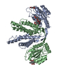 4g86S S: Starting model for refinement |
|---|---|
| Similar structure data |


-Links
 PDBj
PDBj

- Assembly
| Deposited unit | 
| ||||||||
|---|---|---|---|---|---|---|---|---|---|
| 1 |
| ||||||||
| Unit cell |
|
-Components
| #1: Protein | Mass: 33495.066 Da / Num. of mol.: 2 Source method: isolated from a genetically manipulated source Source: (gene. exp.) Synechococcus elongatus (strain PCC 7942) (bacteria)Strain: PCC 7942 / Gene: kaiA, Synpcc7942_1218, see0009 / Plasmid: PET32A+ / Production host: ESCHERICHIA COLI / Strain (production host): BL21 / References: UniProt: Q79PF6 #2: Protein/peptide | Mass: 2247.463 Da / Num. of mol.: 2 / Source method: obtained synthetically / Source: (synth.) synthetic construct (others) / References: UniProt: Q79PF4*PLUS #3: Chemical |   Mass: 407.396 Da / Num. of mol.: 2 / Source method: obtained synthetically / Formula: C21H13NO6S Mass: 407.396 Da / Num. of mol.: 2 / Source method: obtained synthetically / Formula: C21H13NO6S#4: Water | ChemComp-HOH / | Water Mass: 18.015 Da / Num. of mol.: 98 / Source method: isolated from a natural source / Formula: H2O Mass: 18.015 Da / Num. of mol.: 98 / Source method: isolated from a natural source / Formula: H2O |
|---|
-Experimental details
-Experiment
| Experiment | Method: X-RAY DIFFRACTION / Number of used crystals: 1 |
|---|
- Sample preparation
| Crystal | Density Matthews: 2.1 Å3/Da / Density % sol: 41.52 % |
|---|---|
| Crystal grow | Temperature: 289 K / Method: vapor diffusion, hanging drop / pH: 6.8 Details: 180 mM ammonium sulfate, 85 mM sodium cacodylate pH 6.8, 20% PEG 8000 and 15% glycerol in the reservoir. PH range: 6.8 |
-Data collection
| Diffraction | Mean temperature: 100 K |
|---|---|
| Diffraction source | Source: SYNCHROTRON / Site: APS / Beamline: 21-ID-F / Wavelength: 0.978 Å |
| Detector | Type: RAYONIX MX225HE / Detector: CCD / Date: Jun 4, 2014 |
| Radiation | Protocol: SINGLE WAVELENGTH / Monochromatic (M) / Laue (L): M / Scattering type: x-ray |
| Radiation wavelength | Wavelength: 0.978 Å / Relative weight: 1 |
| Reflection | Resolution: 2.82→46.192 Å / Num. obs: 15075 / % possible obs: 100 % / Redundancy: 22 % / Rmerge(I) obs: 0.1 / Net I/av σ(I): 37.1 / Net I/σ(I): 37.1 |
| Reflection shell | Resolution: 2.82→2.89 Å / Redundancy: 22.5 % / Rmerge(I) obs: 0.805 / Mean I/σ(I) obs: 4.7 / % possible all: 100 |
- Processing
| Software |
| ||||||||||||||||||||||||||||||||||||||||||
|---|---|---|---|---|---|---|---|---|---|---|---|---|---|---|---|---|---|---|---|---|---|---|---|---|---|---|---|---|---|---|---|---|---|---|---|---|---|---|---|---|---|---|---|
| Refinement | Method to determine structure: MOLECULAR REPLACEMENT Starting model: 4G86 Resolution: 2.82→46.192 Å / SU ML: 0.39 / Cross valid method: FREE R-VALUE / σ(F): 1.35 / Phase error: 30.96 / Stereochemistry target values: ML
| ||||||||||||||||||||||||||||||||||||||||||
| Solvent computation | Shrinkage radii: 0.9 Å / VDW probe radii: 1.11 Å / Solvent model: FLAT BULK SOLVENT MODEL | ||||||||||||||||||||||||||||||||||||||||||
| Refinement step | Cycle: LAST / Resolution: 2.82→46.192 Å
| ||||||||||||||||||||||||||||||||||||||||||
| Refine LS restraints |
| ||||||||||||||||||||||||||||||||||||||||||
| LS refinement shell |
|
