 Movie
Movie Controller

[English] 日本語
 Yorodumi
- PDB-5c1s: Crystal structure of the GDP-bound fast hydrolyzing mutant (V71A/... -
+ Open data

- Basic information
| Entry | Database: PDB / ID: 5c1s | |||||||||
|---|---|---|---|---|---|---|---|---|---|---|
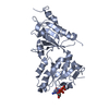
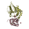
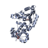
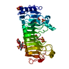
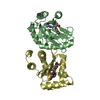
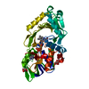
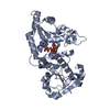
| Title | Crystal structure of the GDP-bound fast hydrolyzing mutant (V71A/K73Q) of EhRabX3 from Entamoeba histolytica | |||||||||
 Components Components | Small GTPase EhRabX3 | |||||||||
 Keywords Keywords |  HYDROLASE / P-loop containing nucleotide triphosphate hydrolases fold / Tandem GTPase HYDROLASE / P-loop containing nucleotide triphosphate hydrolases fold / Tandem GTPase | |||||||||
| Function / homology |  Function and homology information Function and homology information | |||||||||
| Biological species |   Entamoeba histolytica (eukaryote) Entamoeba histolytica (eukaryote) | |||||||||
| Method | X-RAY DIFFRACTION / SYNCHROTRON / SAD / Resolution: 3.1 Å | |||||||||
 Authors Authors | Srivastava, V.K. / Chandra, M. / Datta, S. | |||||||||
| Funding support |  India, 2items India, 2items
| |||||||||
 Citation Citation | Journal: J.Mol.Biol. / Year: 2016 Title: Crystal Structure Analysis of Wild Type and Fast Hydrolyzing Mutant of EhRabX3, a Tandem Ras Superfamily GTPase from Entamoeba histolytica. Authors: Srivastava, V.K. / Chandra, M. / Saito-Nakano, Y. / Nozaki, T. / Datta, S. | |||||||||
| History |
|
- Structure visualization
| Structure viewer | Molecule: MolmilJmol/JSmol |
|---|
- Downloads & links
-Download
| PDBx/mmCIF format | 5c1s.cif.gz | 123 KB | Display | PDBx/mmCIF format |
|---|---|---|---|---|
| PDB format | pdb5c1s.ent.gz | 96.4 KB | Display | PDB format |
| PDBx/mmJSON format | 5c1s.json.gz | Tree view | PDBx/mmJSON format | |
| Others |  Other downloads Other downloads |
-Validation report
| Arichive directory | https://data.pdbj.org/pub/pdb/validation_reports/c1/5c1sftp://data.pdbj.org/pub/pdb/validation_reports/c1/5c1s | HTTPS FTP |
|---|
-Related structure data


-Links
 PDBj
PDBj

- Assembly
| Deposited unit | 
| ||||||||
|---|---|---|---|---|---|---|---|---|---|
| 1 | 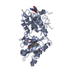
| ||||||||
| 2 | 
| ||||||||
| Unit cell |
|
-Components
| #1: Protein | Mass: 45754.727 Da / Num. of mol.: 2 / Mutation: V71A, K73Q Source method: isolated from a genetically manipulated source Source: (gene. exp.) Entamoeba histolytica (eukaryote) / Gene: EhRabX3 / Plasmid: pET-28a (+) / Production host:  Escherichia coli (E. coli) / Strain (production host): BL21(DE3) / References: UniProt: Q5NT25 Escherichia coli (E. coli) / Strain (production host): BL21(DE3) / References: UniProt: Q5NT25#2: Chemical | Guanosine diphosphate  Type: RNA linking / Mass: 443.201 Da / Num. of mol.: 2 / Source method: obtained synthetically / Formula: C10H15N5O11P2 / Comment: GDP, energy-carrying molecule*YM Type: RNA linking / Mass: 443.201 Da / Num. of mol.: 2 / Source method: obtained synthetically / Formula: C10H15N5O11P2 / Comment: GDP, energy-carrying molecule*YM#3: Water | ChemComp-HOH / | Water Mass: 18.015 Da / Num. of mol.: 54 / Source method: isolated from a natural source / Formula: H2O Mass: 18.015 Da / Num. of mol.: 54 / Source method: isolated from a natural source / Formula: H2O |
|---|
-Experimental details
-Experiment
| Experiment | Method: X-RAY DIFFRACTION |
|---|
- Sample preparation
| Crystal | Density Matthews: 2.11 Å3/Da / Density % sol: 41.74 % |
|---|---|
| Crystal grow | Temperature: 291 K / Method: vapor diffusion, hanging drop / pH: 7 / Details: 200 mM Ammonium citrate, 20% PEG 3350 |
-Data collection
| Diffraction | Mean temperature: 100 K |
|---|---|
| Diffraction source | Source: SYNCHROTRON / Site: ESRF  / Beamline: BM14 / Wavelength: 0.97 Å / Beamline: BM14 / Wavelength: 0.97 Å |
| Detector | Type: MAR CCD 165 mm / Detector: CCD / Date: Nov 13, 2014 |
| Radiation | Protocol: SINGLE WAVELENGTH / Monochromatic (M) / Laue (L): M / Scattering type: x-ray |
| Radiation wavelength | Wavelength: 0.97 Å / Relative weight: 1 |
| Reflection | Resolution: 3.1→38.624 Å / Num. obs: 13133 / % possible obs: 97.5 % / Redundancy: 25.6 % / Net I/σ(I): 20 |
- Processing
| Software |
| ||||||||||||||||||||||||||||||||||||||||||
|---|---|---|---|---|---|---|---|---|---|---|---|---|---|---|---|---|---|---|---|---|---|---|---|---|---|---|---|---|---|---|---|---|---|---|---|---|---|---|---|---|---|---|---|
| Refinement | Method to determine structure: SAD / Resolution: 3.1→38.62 Å / Cross valid method: FREE R-VALUE / σ(F): 1.34 / Phase error: 40.38 / Stereochemistry target values: TWIN_LSQ_F
| ||||||||||||||||||||||||||||||||||||||||||
| Solvent computation | Shrinkage radii: 0.9 Å / VDW probe radii: 1.11 Å / Solvent model: FLAT BULK SOLVENT MODEL | ||||||||||||||||||||||||||||||||||||||||||
| Refinement step | Cycle: LAST / Resolution: 3.1→38.62 Å
| ||||||||||||||||||||||||||||||||||||||||||
| Refine LS restraints |
| ||||||||||||||||||||||||||||||||||||||||||
| LS refinement shell |
|
