 Movie
Movie Controller


+ Open data

- Basic information
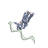
| Entry | Database: PDB / ID: 5c14 | ||||||
|---|---|---|---|---|---|---|---|
| Title | Crystal structure of PECAM-1 D1D2 domain | ||||||
 Components Components | (Platelet endothelial cell adhesion ...) x 2 | ||||||
 Keywords Keywords |  CELL ADHESION / Immunoglobulin / cell adhesion molecule CELL ADHESION / Immunoglobulin / cell adhesion molecule | ||||||
| Function / homology |  Function and homology information Function and homology informationpositive regulation of protein localization to cell-cell junction / diapedesis / cell recognition / glomerular endothelium development / monocyte extravasation / neutrophil extravasation / platelet alpha granule membrane / cell-cell adhesion via plasma-membrane adhesion molecules / bicellular tight junction assembly / negative regulation of immune response ...positive regulation of protein localization to cell-cell junction / diapedesis / cell recognition / glomerular endothelium development / monocyte extravasation / neutrophil extravasation / platelet alpha granule membrane / cell-cell adhesion via plasma-membrane adhesion molecules / bicellular tight junction assembly / negative regulation of immune response / establishment of endothelial barrier / leukocyte cell-cell adhesion / maintenance of blood-brain barrier / Platelet sensitization by LDL / homophilic cell adhesion via plasma membrane adhesion molecules / PECAM1 interactions / Integrin cell surface interactions / phagocytosis / secretory granule membrane / Cell surface interactions at the vascular wall / cell-cell adhesion / transmembrane signaling receptor activity / positive regulation of peptidyl-tyrosine phosphorylation / cell-cell junction / Platelet degranulation / cell surface receptor signaling pathway / positive regulation of cell migration / immune response / membrane raft / positive regulation of protein phosphorylation / external side of plasma membrane / Neutrophil degranulation / signal transduction / protein homodimerization activity / protein-containing complex / extracellular space / extracellular exosome / plasma membraneSimilarity search - Function | ||||||
| Biological species |  Homo sapiens (human) Homo sapiens (human) | ||||||
| Method | X-RAY DIFFRACTION / SYNCHROTRON / SAD / Resolution: 2.8 Å | ||||||
 Authors Authors | Zhou, D. / Paddock, C. / Newman, P. / Zhu, J. | ||||||
 Citation Citation | Journal: Blood / Year: 2016 Title: Structural basis for PECAM-1 homophilic binding. Authors: Paddock, C. / Zhou, D. / Lertkiatmongkol, P. / Newman, P.J. / Zhu, J. | ||||||
| History |
|
- Structure visualization
| Structure viewer | Molecule: MolmilJmol/JSmol |
|---|
- Downloads & links
-Download
| PDBx/mmCIF format | 5c14.cif.gz | 179.5 KB | Display | PDBx/mmCIF format |
|---|---|---|---|---|
| PDB format | pdb5c14.ent.gz | 147.8 KB | Display | PDB format |
| PDBx/mmJSON format | 5c14.json.gz | Tree view | PDBx/mmJSON format | |
| Others |  Other downloads Other downloads |
-Validation report
| Arichive directory | https://data.pdbj.org/pub/pdb/validation_reports/c1/5c14ftp://data.pdbj.org/pub/pdb/validation_reports/c1/5c14 | HTTPS FTP |
|---|
-Related structure data
| Similar structure data |
|---|


-Links
 PDBj
PDBj


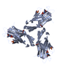
- Assembly
| Deposited unit | 
| ||||||||
|---|---|---|---|---|---|---|---|---|---|
| 1 | 
| ||||||||
| 2 | 
| ||||||||
| 3 | 
| ||||||||
| Unit cell |
|
-Components
-Platelet endothelial cell adhesion ... , 2 types, 2 molecules AB
| #1: Protein | Mass: 24533.736 Da / Num. of mol.: 1 / Fragment: D1D2 domain (UNP residues 28-229) Source method: isolated from a genetically manipulated source Source: (gene. exp.) Homo sapiens (human) / Gene: PECAM1 / Production host:  Drosophila (fruit flies) / References: UniProt: P16284 Drosophila (fruit flies) / References: UniProt: P16284 |
|---|---|
| #2: Protein | Mass: 24486.842 Da / Num. of mol.: 1 / Fragment: D1D2 domain (UNP residues 28-229) Source method: isolated from a genetically manipulated source Source: (gene. exp.) Homo sapiens (human) / Gene: PECAM1 / Production host: Drosophila (fruit flies) / References: UniProt: P16284 |
-Sugars , 1 types, 5 molecules 
| #5: Sugar | ChemComp-NAG / N-Acetylglucosamine Type: D-saccharide, beta linking / Mass: 221.208 Da / Num. of mol.: 5 Type: D-saccharide, beta linking / Mass: 221.208 Da / Num. of mol.: 5Source method: isolated from a genetically manipulated source Formula: C8H15NO6 |
|---|
-Non-polymers , 4 types, 34 molecules 


| #3: Chemical | ChemComp-GOL / Glycerol Mass: 92.094 Da / Num. of mol.: 5 / Source method: obtained synthetically / Formula: C3H8O3 Mass: 92.094 Da / Num. of mol.: 5 / Source method: obtained synthetically / Formula: C3H8O3#4: Chemical | ChemComp-CU / | Copper Mass: 63.546 Da / Num. of mol.: 1 / Source method: obtained synthetically / Formula: Cu Mass: 63.546 Da / Num. of mol.: 1 / Source method: obtained synthetically / Formula: Cu#6: Chemical | ChemComp-TRS / | Tris Mass: 122.143 Da / Num. of mol.: 1 / Source method: obtained synthetically / Formula: C4H12NO3 / Comment: pH buffer*YM Mass: 122.143 Da / Num. of mol.: 1 / Source method: obtained synthetically / Formula: C4H12NO3 / Comment: pH buffer*YM#7: Water | ChemComp-HOH / | WaterMass: 18.015 Da / Num. of mol.: 27 / Source method: isolated from a natural source / Formula: H2O |
|---|
-Experimental details
-Experiment
| Experiment | Method: X-RAY DIFFRACTION / Number of used crystals: 1 |
|---|
- Sample preparation
| Crystal | Density Matthews: 3.89 Å3/Da / Density % sol: 68.36 % |
|---|---|
| Crystal grow | Temperature: 292 K / Method: vapor diffusion, hanging drop / pH: 8.5 / Details: 0.1 M Tris pH 8.5, 0.1 M MgNO3, 20% PEG 3350 |
-Data collection
| Diffraction | Mean temperature: 100 K | |||||||||
|---|---|---|---|---|---|---|---|---|---|---|
| Diffraction source | Source: SYNCHROTRON / Site: APS  / Beamline: 21-ID-D / Wavelength: 0.97624, 0.97872 / Beamline: 21-ID-D / Wavelength: 0.97624, 0.97872 | |||||||||
| Detector | Type: MARMOSAIC 300 mm CCD / Detector: CCD / Date: Nov 1, 2014 | |||||||||
| Radiation | Protocol: MAD / Monochromatic (M) / Laue (L): M / Scattering type: x-ray | |||||||||
| Radiation wavelength |
| |||||||||
| Reflection | Resolution: 2.8→20 Å / Num. obs: 36261 / % possible obs: 99.6 % / Redundancy: 6.17 % / Rmerge(I) obs: 0.125 / Net I/σ(I): 11.76 | |||||||||
| Reflection shell | Resolution: 2.8→3.2 Å / Redundancy: 4.79 % / Rmerge(I) obs: 0.493 / Mean I/σ(I) obs: 2.42 / Num. unique all: 11969 / % possible all: 99.8 |
- Processing
| Software |
| ||||||||||||||||||||||||||||||||||||||||||||||||||||||||
|---|---|---|---|---|---|---|---|---|---|---|---|---|---|---|---|---|---|---|---|---|---|---|---|---|---|---|---|---|---|---|---|---|---|---|---|---|---|---|---|---|---|---|---|---|---|---|---|---|---|---|---|---|---|---|---|---|---|
| Refinement | Method to determine structure: SAD / Resolution: 2.8→19.79 Å / SU ML: 0.71 / Cross valid method: FREE R-VALUE / σ(F): 1.45 / Phase error: 36.67 / Stereochemistry target values: ML
| ||||||||||||||||||||||||||||||||||||||||||||||||||||||||
| Solvent computation | Shrinkage radii: 0.9 Å / VDW probe radii: 1.11 Å / Solvent model: FLAT BULK SOLVENT MODEL | ||||||||||||||||||||||||||||||||||||||||||||||||||||||||
| Refinement step | Cycle: LAST / Resolution: 2.8→19.79 Å
| ||||||||||||||||||||||||||||||||||||||||||||||||||||||||
| Refine LS restraints |
| ||||||||||||||||||||||||||||||||||||||||||||||||||||||||
| LS refinement shell |
| ||||||||||||||||||||||||||||||||||||||||||||||||||||||||
| Refinement TLS params. | Method: refined / Origin x: 19.9177 Å / Origin y: 33.0702 Å / Origin z: -0.8912 Å
| ||||||||||||||||||||||||||||||||||||||||||||||||||||||||
| Refinement TLS group | Selection details: ALL |
