 Movie
Movie Controller


+ Open data

- Basic information
| Entry | Database: PDB / ID: 5bza | ||||||
|---|---|---|---|---|---|---|---|
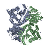
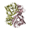
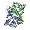
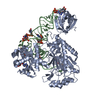
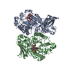
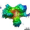
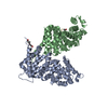
| Title | Crystal structure of CbsA from Thermotoga neapolitana | ||||||
 Components Components | Beta-N-acetylhexosaminidase Hexosaminidase Hexosaminidase | ||||||
 Keywords Keywords | HYDROLASE / cbsA / Thermotoga / thermostable enzyme / beta-N-acetylglucosaminidase | ||||||
| Function / homology |  Function and homology information Function and homology informationhydrolase activity, hydrolyzing O-glycosyl compounds / carbohydrate metabolic process / metal ion bindingSimilarity search - Function | ||||||
| Biological species |   Thermotoga neapolitana (bacteria) Thermotoga neapolitana (bacteria) | ||||||
| Method | X-RAY DIFFRACTION / SYNCHROTRON / Resolution: 2.002 Å | ||||||
 Authors Authors | Ha, N.C. / Kim, J.S. / Yoon, B.Y. | ||||||
 Citation Citation | Journal: Biochem.Biophys.Res.Commun. / Year: 2015 Title: Crystal structure of beta-N-acetylglucosaminidase CbsA from Thermotoga neapolitana Authors: Kim, J.S. / Yoon, B.Y. / Ahn, J. / Cha, J. / Ha, N.C. #1: Journal: Acta Crystallogr.,Sect.F / Year: 2012 Title: Crystallization and preliminary X-ray crystallographic analysis of the beta-N-acetylglucosaminidase CbsA from Thermotoga neapolitana Authors: Yoon, B.Y. / Jiao, L. / Moon, H.R. / Cha, J. / Ha, N.C. | ||||||
| History |
|
- Structure visualization
| Structure viewer | Molecule: MolmilJmol/JSmol |
|---|
- Downloads & links
-Download
| PDBx/mmCIF format | 5bza.cif.gz | 362.4 KB | Display | PDBx/mmCIF format |
|---|---|---|---|---|
| PDB format | pdb5bza.ent.gz | 305.4 KB | Display | PDB format |
| PDBx/mmJSON format | 5bza.json.gz | Tree view | PDBx/mmJSON format | |
| Others |  Other downloads Other downloads |
-Validation report
| Arichive directory | https://data.pdbj.org/pub/pdb/validation_reports/bz/5bzaftp://data.pdbj.org/pub/pdb/validation_reports/bz/5bza | HTTPS FTP |
|---|
-Related structure data


-Links
 PDBj
PDBj- Assembly
| Deposited unit | 
| ||||||||
|---|---|---|---|---|---|---|---|---|---|
| 1 | 
| ||||||||
| 2 | 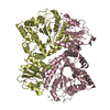
| ||||||||
| Unit cell |
|
-Components
| #1: Protein | Hexosaminidase Mass: 52637.305 Da / Num. of mol.: 4 Source method: isolated from a genetically manipulated source Source: (gene. exp.) Thermotoga neapolitana (bacteria) / Gene: cbsA / Production host: Escherichia coli (E. coli) / References: UniProt: Q9AG27, beta-N-acetylhexosaminidase#2: Chemical | ChemComp-CD /   Mass: 112.411 Da / Num. of mol.: 33 / Source method: obtained synthetically / Formula: Cd Mass: 112.411 Da / Num. of mol.: 33 / Source method: obtained synthetically / Formula: Cd#3: Water | ChemComp-HOH / | Water Mass: 18.015 Da / Num. of mol.: 627 / Source method: isolated from a natural source / Formula: H2O Mass: 18.015 Da / Num. of mol.: 627 / Source method: isolated from a natural source / Formula: H2O |
|---|
-Experimental details
-Experiment
| Experiment | Method: X-RAY DIFFRACTION / Number of used crystals: 1 |
|---|
- Sample preparation
| Crystal | Density Matthews: 2.99 Å3/Da / Density % sol: 58.82 % |
|---|---|
| Crystal grow | Temperature: 287 K / Method: vapor diffusion, hanging drop Details: 0.1 M HEPES pH 7.5 1.0 M sodium acetate 0.05 M CdCl2 |
-Data collection
| Diffraction | Mean temperature: 100 K |
|---|---|
| Diffraction source | Source: SYNCHROTRON / Site: SPring-8  / Beamline: BL44B2 / Wavelength: 0.9 Å / Beamline: BL44B2 / Wavelength: 0.9 Å |
| Detector | Type: MAR CCD 165 mm / Detector: CCD / Date: Jun 30, 2011 |
| Radiation | Protocol: SINGLE WAVELENGTH / Monochromatic (M) / Laue (L): M / Scattering type: x-ray |
| Radiation wavelength | Wavelength: 0.9 Å / Relative weight: 1 |
| Reflection | Resolution: 2→50 Å / Num. obs: 158414 / % possible obs: 94.2 % / Redundancy: 4.5 % / Net I/σ(I): 9.25 |
- Processing
| Software |
| |||||||||||||||||||||||||||||||||||||||||||||||||||||||||||||||||||||||||||||||||||||||||||||||||||||||||
|---|---|---|---|---|---|---|---|---|---|---|---|---|---|---|---|---|---|---|---|---|---|---|---|---|---|---|---|---|---|---|---|---|---|---|---|---|---|---|---|---|---|---|---|---|---|---|---|---|---|---|---|---|---|---|---|---|---|---|---|---|---|---|---|---|---|---|---|---|---|---|---|---|---|---|---|---|---|---|---|---|---|---|---|---|---|---|---|---|---|---|---|---|---|---|---|---|---|---|---|---|---|---|---|---|---|---|
| Refinement | Resolution: 2.002→39.741 Å / SU ML: 0.37 / Cross valid method: FREE R-VALUE / σ(F): 1.44 / Phase error: 31.23 / Stereochemistry target values: ML
| |||||||||||||||||||||||||||||||||||||||||||||||||||||||||||||||||||||||||||||||||||||||||||||||||||||||||
| Solvent computation | Shrinkage radii: 0.9 Å / VDW probe radii: 1.11 Å / Solvent model: FLAT BULK SOLVENT MODEL | |||||||||||||||||||||||||||||||||||||||||||||||||||||||||||||||||||||||||||||||||||||||||||||||||||||||||
| Refinement step | Cycle: LAST / Resolution: 2.002→39.741 Å
| |||||||||||||||||||||||||||||||||||||||||||||||||||||||||||||||||||||||||||||||||||||||||||||||||||||||||
| Refine LS restraints |
| |||||||||||||||||||||||||||||||||||||||||||||||||||||||||||||||||||||||||||||||||||||||||||||||||||||||||
| LS refinement shell |
|
