 Movie
Movie Controller

[English] 日本語
 Yorodumi
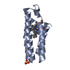
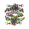
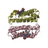
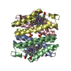
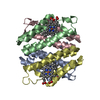
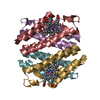
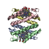
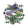
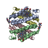
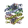
- PDB-5bu7: Crystal structure of an engineered protein that forms nanotubes w... -
+ Open data

- Basic information
| Entry | Database: PDB / ID: 5bu7 | ||||||
|---|---|---|---|---|---|---|---|
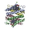
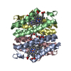
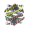
| Title | Crystal structure of an engineered protein that forms nanotubes with tunable diameters | ||||||
 Components Components | Soluble cytochrome b562 | ||||||
 Keywords Keywords | METAL BINDING PROTEIN /  Metalloprotein / Hemeprotein Metalloprotein / Hemeprotein | ||||||
| Function / homology |  Function and homology informationelectron transport chain / periplasmic space / electron transfer activity / iron ion binding / heme binding Function and homology informationelectron transport chain / periplasmic space / electron transfer activity / iron ion binding / heme bindingSimilarity search - Function | ||||||
| Biological species |  Escherichia coli (E. coli) Escherichia coli (E. coli) | ||||||
| Method | X-RAY DIFFRACTION / SYNCHROTRON / MOLECULAR REPLACEMENT / Resolution: 2.46 Å | ||||||
 Authors Authors | Brodin, J.D. / Smith, S.J. / Tezcan, F.A. | ||||||
| Funding support |  United States, 1items United States, 1items
| ||||||
 Citation Citation | Journal: To be Published Title: Designed, Helical Protein Nanotubes with Tunable Diameters from a Single Building Block. Authors: Brodin, J.D. / Smith, S.J. / Tezcan, F.A. | ||||||
| History |
|
- Structure visualization
| Structure viewer | Molecule: MolmilJmol/JSmol |
|---|
- Downloads & links
-Download
| PDBx/mmCIF format | 5bu7.cif.gz | 55.7 KB | Display | PDBx/mmCIF format |
|---|---|---|---|---|
| PDB format | pdb5bu7.ent.gz | 43.4 KB | Display | PDB format |
| PDBx/mmJSON format | 5bu7.json.gz | Tree view | PDBx/mmJSON format | |
| Others |  Other downloads Other downloads |
-Validation report
| Arichive directory | https://data.pdbj.org/pub/pdb/validation_reports/bu/5bu7ftp://data.pdbj.org/pub/pdb/validation_reports/bu/5bu7 | HTTPS FTP |
|---|
-Related structure data
| Related structure data | |
|---|---|
| Similar structure data |
-Links
 PDBj
PDBj


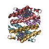
- Assembly
| Deposited unit | 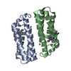
| |||||||||||||||||||||||||||
|---|---|---|---|---|---|---|---|---|---|---|---|---|---|---|---|---|---|---|---|---|---|---|---|---|---|---|---|---|
| 1 | 
| |||||||||||||||||||||||||||
| Unit cell |
| |||||||||||||||||||||||||||
| Components on special symmetry positions |
| |||||||||||||||||||||||||||
| Noncrystallographic symmetry (NCS) | NCS domain:
NCS domain segments:
|
-Components
| #1: Protein | Mass: 11615.114 Da / Num. of mol.: 2 Mutation: T96C, D73H, K77H, R98C, Y101C, K27E, D28K, T31E, R34L, L38A, Q41L, H59R, D66A, V69M, L76A Source method: isolated from a genetically manipulated source Source: (gene. exp.) Escherichia coli (E. coli) / Gene: cybC / Production host: Escherichia coli (E. coli) / References: UniProt: P0ABE7#2: Chemical | Heme B  Mass: 616.487 Da / Num. of mol.: 2 / Source method: obtained synthetically / Formula: C34H32FeN4O4 Mass: 616.487 Da / Num. of mol.: 2 / Source method: obtained synthetically / Formula: C34H32FeN4O4#3: Chemical | ChemComp-ZN /   Mass: 65.409 Da / Num. of mol.: 7 / Source method: obtained synthetically / Formula: Zn Mass: 65.409 Da / Num. of mol.: 7 / Source method: obtained synthetically / Formula: Zn#4: Water | ChemComp-HOH / | Water Mass: 18.015 Da / Num. of mol.: 56 / Source method: isolated from a natural source / Formula: H2O Mass: 18.015 Da / Num. of mol.: 56 / Source method: isolated from a natural source / Formula: H2O |
|---|
-Experimental details
-Experiment
| Experiment | Method: X-RAY DIFFRACTION / Number of used crystals: 1 |
|---|
- Sample preparation
| Crystal | Density Matthews: 2.23 Å3/Da / Density % sol: 44.95 % |
|---|---|
| Crystal grow | Temperature: 298 K / Method: batch mode / pH: 7.5 / Details: 50-fold excess of zinc chloride |
-Data collection
| Diffraction | Mean temperature: 80 K |
|---|---|
| Diffraction source | Source: SYNCHROTRON / Site: SSRL / Beamline: BL12-2 / Wavelength: 0.98 Å |
| Detector | Type: DECTRIS PILATUS 6M / Detector: PIXEL / Date: Jan 22, 2013 |
| Radiation | Monochromator: double crystal Si(111) / Protocol: SINGLE WAVELENGTH / Monochromatic (M) / Laue (L): M / Scattering type: x-ray |
| Radiation wavelength | Wavelength: 0.98 Å / Relative weight: 1 |
| Reflection | Resolution: 2.46→85.69 Å / Num. obs: 8104 / % possible obs: 95.1 % / Redundancy: 3.5 % / Net I/σ(I): 6.7 |
- Processing
| Software |
| ||||||||||||||||||||||||||||||||||||||||||||||||||||||||||||
|---|---|---|---|---|---|---|---|---|---|---|---|---|---|---|---|---|---|---|---|---|---|---|---|---|---|---|---|---|---|---|---|---|---|---|---|---|---|---|---|---|---|---|---|---|---|---|---|---|---|---|---|---|---|---|---|---|---|---|---|---|---|
| Refinement | Method to determine structure: MOLECULAR REPLACEMENT / Resolution: 2.46→45.8 Å / Cor.coef. Fo:Fc: 0.934 / Cor.coef. Fo:Fc free: 0.887 / WRfactor Rfree: 0.3314 / WRfactor Rwork: 0.2528 / FOM work R set: 0.773 / SU B: 13.14 / SU ML: 0.288 / SU R Cruickshank DPI: 1.152 / SU Rfree: 0.3638 / Cross valid method: THROUGHOUT / σ(F): 0 / ESU R: 1.152 / ESU R Free: 0.364 / Stereochemistry target values: MAXIMUM LIKELIHOOD / Details: HYDROGENS HAVE BEEN ADDED IN THE RIDING POSITIONS
| ||||||||||||||||||||||||||||||||||||||||||||||||||||||||||||
| Solvent computation | Ion probe radii: 0.8 Å / Shrinkage radii: 0.8 Å / VDW probe radii: 1.2 Å / Solvent model: MASK | ||||||||||||||||||||||||||||||||||||||||||||||||||||||||||||
| Displacement parameters | Biso max: 107.19 Å2 / Biso mean: 55.873 Å2 / Biso min: 23.69 Å2
| ||||||||||||||||||||||||||||||||||||||||||||||||||||||||||||
| Refinement step | Cycle: final / Resolution: 2.46→45.8 Å
| ||||||||||||||||||||||||||||||||||||||||||||||||||||||||||||
| Refine LS restraints |
| ||||||||||||||||||||||||||||||||||||||||||||||||||||||||||||
| Refine LS restraints NCS | Ens-ID: 1 / Number: 6045 / Refine-ID: X-RAY DIFFRACTION / Type: interatomic distance / Rms dev position: 0.08 Å / Weight position: 0.05
| ||||||||||||||||||||||||||||||||||||||||||||||||||||||||||||
| LS refinement shell | Resolution: 2.461→2.525 Å / Total num. of bins used: 20
|
