 Movie
Movie Controller

[English] 日本語
 Yorodumi
- PDB-5bmw: Crystal structure of T75V mutant of Triosephosphate isomerase fro... -
+ Open data

- Basic information
| Entry | Database: PDB / ID: 5bmw | ||||||
|---|---|---|---|---|---|---|---|
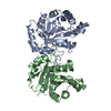
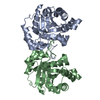
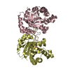
| Title | Crystal structure of T75V mutant of Triosephosphate isomerase from Plasmodium falciparum | ||||||
 Components Components | Triosephosphate isomerase | ||||||
 Keywords Keywords | ISOMERASE / TIM Barrel | ||||||
| Function / homology |  Function and homology informationtriose-phosphate isomerase / triose-phosphate isomerase activity / gluconeogenesis / glycolytic process / identical protein binding Function and homology informationtriose-phosphate isomerase / triose-phosphate isomerase activity / gluconeogenesis / glycolytic process / identical protein bindingSimilarity search - Function | ||||||
| Biological species |  Plasmodium falciparum (malaria parasite P. falciparum) Plasmodium falciparum (malaria parasite P. falciparum) | ||||||
| Method | X-RAY DIFFRACTION / MOLECULAR REPLACEMENT / Resolution: 1.86 Å | ||||||
 Authors Authors | Bandyopadhyay, D. / Murthy, M.R.N. / Balaram, H. / Balaram, P. | ||||||
| Funding support |  India, 1items India, 1items
| ||||||
 Citation Citation | Journal: Febs J. / Year: 2015 Title: Probing the role of highly conserved residues in triosephosphate isomerase - analysis of site specific mutants at positions 64 and 75 in the Plasmodial enzyme Authors: Bandyopadhyay, D. / Murthy, M.R. / Balaram, H. / Balaram, P. | ||||||
| History |
|
- Structure visualization
| Structure viewer | Molecule: MolmilJmol/JSmol |
|---|
- Downloads & links
-Download
| PDBx/mmCIF format | 5bmw.cif.gz | 123 KB | Display | PDBx/mmCIF format |
|---|---|---|---|---|
| PDB format | pdb5bmw.ent.gz | 92.5 KB | Display | PDB format |
| PDBx/mmJSON format | 5bmw.json.gz | Tree view | PDBx/mmJSON format | |
| Others |  Other downloads Other downloads |
-Validation report
| Arichive directory | https://data.pdbj.org/pub/pdb/validation_reports/bm/5bmwftp://data.pdbj.org/pub/pdb/validation_reports/bm/5bmw | HTTPS FTP |
|---|
-Related structure data
| Related structure data |  4zz9C  5bmxC  5bnkC 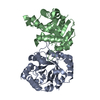 5brbC  1o5xS S: Starting model for refinement C: citing same article ( |
|---|---|
| Similar structure data |


-Links
 PDBj
PDBj
- Assembly
| Deposited unit | 
| ||||||||
|---|---|---|---|---|---|---|---|---|---|
| 1 |
| ||||||||
| Unit cell |
|
-Components
-Protein , 1 types, 2 molecules AB
| #1: Protein | / TIM / Triose-phosphate isomerase Mass: 27995.766 Da / Num. of mol.: 2 / Mutation: T75V, A163V Source method: isolated from a genetically manipulated source Source: (gene. exp.) Plasmodium falciparum (malaria parasite P. falciparum)Gene: TPI / Plasmid: pTrc99A / Production host:  ESCHERICHIA COLI (E. coli) / Strain (production host): AA200 / References: UniProt: Q07412, triose-phosphate isomerase ESCHERICHIA COLI (E. coli) / Strain (production host): AA200 / References: UniProt: Q07412, triose-phosphate isomerase |
|---|
-Non-polymers , 5 types, 495 molecules 


| #2: Chemical | ChemComp-EDO / Ethylene glycol Mass: 62.068 Da / Num. of mol.: 8 / Source method: obtained synthetically / Formula: C2H6O2 Mass: 62.068 Da / Num. of mol.: 8 / Source method: obtained synthetically / Formula: C2H6O2#3: Chemical |  Mass: 22.990 Da / Num. of mol.: 2 / Source method: obtained synthetically / Formula: Na Mass: 22.990 Da / Num. of mol.: 2 / Source method: obtained synthetically / Formula: Na#4: Chemical | ChemComp-MES / | MES (buffer) Mass: 195.237 Da / Num. of mol.: 1 / Source method: obtained synthetically / Formula: C6H13NO4S / Comment: pH buffer*YM Mass: 195.237 Da / Num. of mol.: 1 / Source method: obtained synthetically / Formula: C6H13NO4S / Comment: pH buffer*YM#5: Chemical | ChemComp-CA / |  Mass: 40.078 Da / Num. of mol.: 1 / Source method: obtained synthetically / Formula: Ca Mass: 40.078 Da / Num. of mol.: 1 / Source method: obtained synthetically / Formula: Ca#6: Water | ChemComp-HOH / | WaterMass: 18.015 Da / Num. of mol.: 483 / Source method: isolated from a natural source / Formula: H2O |
|---|
-Experimental details
-Experiment
| Experiment | Method: X-RAY DIFFRACTION / Number of used crystals: 1 |
|---|
- Sample preparation
| Crystal | Density Matthews: 1.97 Å3/Da / Density % sol: 37.62 % |
|---|---|
| Crystal grow | Temperature: 296 K / Method: vapor diffusion, hanging drop / pH: 6.5 Details: 20% PEG 1450, 100 mM MES buffer, 10 mM Calcium chloride, 0.5 mM EDTA, 0.5 mM DTT, 0.5 mM sodium azide |
-Data collection
| Diffraction | Mean temperature: 100 K | ||||||||||||||||||||||||||||||||||||||||||||||||||||||||||||||||||||||||||||||||||||||||||||||||||||||||||||||
|---|---|---|---|---|---|---|---|---|---|---|---|---|---|---|---|---|---|---|---|---|---|---|---|---|---|---|---|---|---|---|---|---|---|---|---|---|---|---|---|---|---|---|---|---|---|---|---|---|---|---|---|---|---|---|---|---|---|---|---|---|---|---|---|---|---|---|---|---|---|---|---|---|---|---|---|---|---|---|---|---|---|---|---|---|---|---|---|---|---|---|---|---|---|---|---|---|---|---|---|---|---|---|---|---|---|---|---|---|---|---|---|
| Diffraction source | Source: ROTATING ANODE / Type: RIGAKU RU200 / Wavelength: 1.541 Å | ||||||||||||||||||||||||||||||||||||||||||||||||||||||||||||||||||||||||||||||||||||||||||||||||||||||||||||||
| Detector | Type: MAR scanner 345 mm plate / Detector: IMAGE PLATE / Date: Apr 10, 2014 | ||||||||||||||||||||||||||||||||||||||||||||||||||||||||||||||||||||||||||||||||||||||||||||||||||||||||||||||
| Radiation | Monochromator: Osmic mirrors / Protocol: SINGLE WAVELENGTH / Monochromatic (M) / Laue (L): M / Scattering type: x-ray | ||||||||||||||||||||||||||||||||||||||||||||||||||||||||||||||||||||||||||||||||||||||||||||||||||||||||||||||
| Radiation wavelength | Wavelength: 1.541 Å / Relative weight: 1 | ||||||||||||||||||||||||||||||||||||||||||||||||||||||||||||||||||||||||||||||||||||||||||||||||||||||||||||||
| Reflection | Resolution: 1.859→73.73 Å / Num. all: 36392 / Num. obs: 36392 / % possible obs: 99.8 % / Redundancy: 5.6 % / Rpim(I) all: 0.025 / Rrim(I) all: 0.061 / Rsym value: 0.056 / Net I/av σ(I): 9.916 / Net I/σ(I): 22.2 / Num. measured all: 203849 | ||||||||||||||||||||||||||||||||||||||||||||||||||||||||||||||||||||||||||||||||||||||||||||||||||||||||||||||
| Reflection shell | Diffraction-ID: 1 / Rejects: 0
|
- Processing
| Software |
| |||||||||||||||||||||||||||||||||||||||||||||||||||||||||||||||||||||||||||
|---|---|---|---|---|---|---|---|---|---|---|---|---|---|---|---|---|---|---|---|---|---|---|---|---|---|---|---|---|---|---|---|---|---|---|---|---|---|---|---|---|---|---|---|---|---|---|---|---|---|---|---|---|---|---|---|---|---|---|---|---|---|---|---|---|---|---|---|---|---|---|---|---|---|---|---|---|
| Refinement | Method to determine structure: MOLECULAR REPLACEMENT Starting model: 1O5X Resolution: 1.86→73.73 Å / Cor.coef. Fo:Fc: 0.964 / Cor.coef. Fo:Fc free: 0.941 / WRfactor Rfree: 0.1829 / WRfactor Rwork: 0.1316 / FOM work R set: 0.9111 / SU B: 2.335 / SU ML: 0.071 / SU R Cruickshank DPI: 0.1271 / SU Rfree: 0.1234 / Cross valid method: THROUGHOUT / σ(F): 0 / ESU R: 0.127 / ESU R Free: 0.123 / Stereochemistry target values: MAXIMUM LIKELIHOOD Details: HYDROGENS HAVE BEEN ADDED IN THE RIDING POSITIONS U VALUES : REFINED INDIVIDUALLY
| |||||||||||||||||||||||||||||||||||||||||||||||||||||||||||||||||||||||||||
| Solvent computation | Ion probe radii: 0.8 Å / Shrinkage radii: 0.8 Å / VDW probe radii: 1.2 Å / Solvent model: MASK | |||||||||||||||||||||||||||||||||||||||||||||||||||||||||||||||||||||||||||
| Displacement parameters | Biso max: 46.58 Å2 / Biso mean: 11.586 Å2 / Biso min: 4.79 Å2
| |||||||||||||||||||||||||||||||||||||||||||||||||||||||||||||||||||||||||||
| Refinement step | Cycle: final / Resolution: 1.86→73.73 Å
| |||||||||||||||||||||||||||||||||||||||||||||||||||||||||||||||||||||||||||
| Refine LS restraints |
| |||||||||||||||||||||||||||||||||||||||||||||||||||||||||||||||||||||||||||
| LS refinement shell | Resolution: 1.859→1.907 Å / Total num. of bins used: 20
|
