 Movie
Movie Controller

[English] 日本語
 Yorodumi
- PDB-5b27: The 1.02A structure of human FABP3 M20S mutant complexed with pal... -
+ Open data

- Basic information
| Entry | Database: PDB / ID: 5b27 | |||||||||||||||
|---|---|---|---|---|---|---|---|---|---|---|---|---|---|---|---|---|
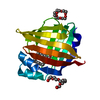
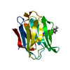
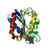
| Title | The 1.02A structure of human FABP3 M20S mutant complexed with palmitic acid | |||||||||||||||
 Components Components | Fatty acid-binding protein, heart | |||||||||||||||
 Keywords Keywords | LIPID BINDING PROTEIN /  complex / palmitic acid complex / palmitic acid | |||||||||||||||
| Function / homology |  Function and homology information Function and homology informationpositive regulation of long-chain fatty acid import into cell / regulation of phosphatidylcholine biosynthetic process / regulation of fatty acid oxidation / positive regulation of phospholipid biosynthetic process / intracellular lipid transport / oleic acid binding / phospholipid homeostasis / long-chain fatty acid binding / Triglyceride catabolism / long-chain fatty acid transport ...positive regulation of long-chain fatty acid import into cell / regulation of phosphatidylcholine biosynthetic process / regulation of fatty acid oxidation / positive regulation of phospholipid biosynthetic process / intracellular lipid transport / oleic acid binding / phospholipid homeostasis / long-chain fatty acid binding / Triglyceride catabolism / long-chain fatty acid transport / brown fat cell differentiation / cytoskeletal protein binding / cholesterol homeostasis / negative regulation of cell population proliferation / extracellular space / extracellular exosome / nucleus / cytosolSimilarity search - Function | |||||||||||||||
| Biological species |  Homo sapiens (human) Homo sapiens (human) | |||||||||||||||
| Method | X-RAY DIFFRACTION / SYNCHROTRON / MOLECULAR REPLACEMENT / Resolution: 1.02 Å | |||||||||||||||
 Authors Authors | Matsuoka, D. / Sugiyama, S. / Kakinouchi, K. / Niiyama, M. / Murata, M. / Matsuoka, S. | |||||||||||||||
| Funding support |  Japan, 4items Japan, 4items
| |||||||||||||||
 Citation Citation | Journal: To Be Published Title: The 1.02A structure of human FABP3 M20S mutant complexed with palmitic acid. Authors: Matsuoka, D. / Sugiyama, S. / Kakinouchi, K. / Niiyama, M. / Murata, M. / Matsuoka, S. | |||||||||||||||
| History |
|
- Structure visualization
| Structure viewer | Molecule: MolmilJmol/JSmol |
|---|
- Downloads & links
-Download
| PDBx/mmCIF format | 5b27.cif.gz | 91.5 KB | Display | PDBx/mmCIF format |
|---|---|---|---|---|
| PDB format | pdb5b27.ent.gz | 68.2 KB | Display | PDB format |
| PDBx/mmJSON format | 5b27.json.gz | Tree view | PDBx/mmJSON format | |
| Others |  Other downloads Other downloads |
-Validation report
| Arichive directory | https://data.pdbj.org/pub/pdb/validation_reports/b2/5b27ftp://data.pdbj.org/pub/pdb/validation_reports/b2/5b27 | HTTPS FTP |
|---|
-Related structure data
| Related structure data | 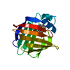 2hmbS S: Starting model for refinement |
|---|---|
| Similar structure data |


-Links
 PDBj
PDBj


- Assembly
| Deposited unit | 
| ||||||||
|---|---|---|---|---|---|---|---|---|---|
| 1 |
| ||||||||
| Unit cell |
|
-Components
| #1: Protein | Mass: 14834.903 Da / Num. of mol.: 1 / Mutation: M20S Source method: isolated from a genetically manipulated source Source: (gene. exp.) Homo sapiens (human) / Gene: FABP3, FABP11, MDGI / Production host:  Escherichia coli (E. coli) / References: UniProt: P05413 Escherichia coli (E. coli) / References: UniProt: P05413 |
|---|---|
| #2: Chemical | ChemComp-PLM / Palmitic acid  Mass: 256.424 Da / Num. of mol.: 1 / Source method: obtained synthetically / Formula: C16H32O2 Mass: 256.424 Da / Num. of mol.: 1 / Source method: obtained synthetically / Formula: C16H32O2 |
| #3: Chemical | ChemComp-PG4 / Polyethylene glycol  Mass: 194.226 Da / Num. of mol.: 1 / Source method: obtained synthetically / Formula: C8H18O5 / Comment: precipitant*YM Mass: 194.226 Da / Num. of mol.: 1 / Source method: obtained synthetically / Formula: C8H18O5 / Comment: precipitant*YM |
| #4: Water | ChemComp-HOH / Water Mass: 18.015 Da / Num. of mol.: 229 / Source method: isolated from a natural source / Formula: H2O Mass: 18.015 Da / Num. of mol.: 229 / Source method: isolated from a natural source / Formula: H2O |
-Experimental details
-Experiment
| Experiment | Method: X-RAY DIFFRACTION |
|---|
- Sample preparation
| Crystal | Density Matthews: 2.19 Å3/Da / Density % sol: 43.9 % |
|---|---|
| Crystal grow | Temperature: 293 K / Method: vapor diffusion, sitting drop / pH: 8 / Details: 0.1M Tris-HCl (pH8.0), 45% (v/v) PEG 400 |
-Data collection
| Diffraction | Mean temperature: 100 K |
|---|---|
| Diffraction source | Source: SYNCHROTRON / Site: SPring-8 / Beamline: BL38B1 / Wavelength: 0.9 Å |
| Detector | Type: ADSC QUANTUM 315 / Detector: CCD / Date: Jan 29, 2013 |
| Radiation | Protocol: SINGLE WAVELENGTH / Monochromatic (M) / Laue (L): M / Scattering type: x-ray |
| Radiation wavelength | Wavelength: 0.9 Å / Relative weight: 1 |
| Reflection | Resolution: 1.02→43.07 Å / Num. obs: 66771 / % possible obs: 99.5 % / Redundancy: 99.5 % / Rmerge(I) obs: 0.09 / Net I/σ(I): 7 |
| Reflection shell | Resolution: 1.02→1.04 Å / Redundancy: 9.3 % / Rmerge(I) obs: 0.37 / Mean I/σ(I) obs: 3.7 / % possible all: 99.9 |
- Processing
| Software |
| ||||||||||||||||||||||||||||||||||||||||||||||||||||||||||||||||||||||||||||||||||||||||||||||||||||||||||||||||||||||||||||||||||||||||||||||||||||||||||||||||||||||||||||||||||||||
|---|---|---|---|---|---|---|---|---|---|---|---|---|---|---|---|---|---|---|---|---|---|---|---|---|---|---|---|---|---|---|---|---|---|---|---|---|---|---|---|---|---|---|---|---|---|---|---|---|---|---|---|---|---|---|---|---|---|---|---|---|---|---|---|---|---|---|---|---|---|---|---|---|---|---|---|---|---|---|---|---|---|---|---|---|---|---|---|---|---|---|---|---|---|---|---|---|---|---|---|---|---|---|---|---|---|---|---|---|---|---|---|---|---|---|---|---|---|---|---|---|---|---|---|---|---|---|---|---|---|---|---|---|---|---|---|---|---|---|---|---|---|---|---|---|---|---|---|---|---|---|---|---|---|---|---|---|---|---|---|---|---|---|---|---|---|---|---|---|---|---|---|---|---|---|---|---|---|---|---|---|---|---|---|
| Refinement | Method to determine structure: MOLECULAR REPLACEMENT Starting model: 2HMB Resolution: 1.02→43.07 Å / Cor.coef. Fo:Fc: 0.981 / Cor.coef. Fo:Fc free: 0.978 / SU B: 0.513 / SU ML: 0.012 / Cross valid method: THROUGHOUT / ESU R: 0.021 / ESU R Free: 0.022 / Stereochemistry target values: MAXIMUM LIKELIHOOD / Details: HYDROGENS HAVE BEEN ADDED IN THE RIDING POSITIONS
| ||||||||||||||||||||||||||||||||||||||||||||||||||||||||||||||||||||||||||||||||||||||||||||||||||||||||||||||||||||||||||||||||||||||||||||||||||||||||||||||||||||||||||||||||||||||
| Solvent computation | Ion probe radii: 0.8 Å / Shrinkage radii: 0.8 Å / VDW probe radii: 1.4 Å / Solvent model: BABINET MODEL WITH MASK | ||||||||||||||||||||||||||||||||||||||||||||||||||||||||||||||||||||||||||||||||||||||||||||||||||||||||||||||||||||||||||||||||||||||||||||||||||||||||||||||||||||||||||||||||||||||
| Displacement parameters | Biso mean: 10.673 Å2
| ||||||||||||||||||||||||||||||||||||||||||||||||||||||||||||||||||||||||||||||||||||||||||||||||||||||||||||||||||||||||||||||||||||||||||||||||||||||||||||||||||||||||||||||||||||||
| Refinement step | Cycle: 1 / Resolution: 1.02→43.07 Å
| ||||||||||||||||||||||||||||||||||||||||||||||||||||||||||||||||||||||||||||||||||||||||||||||||||||||||||||||||||||||||||||||||||||||||||||||||||||||||||||||||||||||||||||||||||||||
| Refine LS restraints |
|