 Movie
Movie Controller

[English] 日本語
 Yorodumi
- PDB-5b1j: Crystal structure of the electron-transfer complex of copper nitr... -
+ Open data

- Basic information
| Entry | Database: PDB / ID: 5b1j | ||||||
|---|---|---|---|---|---|---|---|
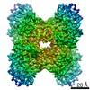
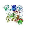
| Title | Crystal structure of the electron-transfer complex of copper nitrite reductase with a cupredoxin | ||||||
 Components Components |
| ||||||
 Keywords Keywords | OXIDOREDUCTASE/ELECTRON TRANSPORT /  complex / copper nitrite reductase / electron transfer / OXIDOREDUCTASE-ELECTRON TRANSPORT complex complex / copper nitrite reductase / electron transfer / OXIDOREDUCTASE-ELECTRON TRANSPORT complex | ||||||
| Function / homology |  Function and homology information Function and homology informationdenitrification pathway / nitrite reductase (NO-forming) / nitrite reductase (NO-forming) activity / nitrate assimilation / periplasmic space / electron transfer activity / copper ion bindingSimilarity search - Function | ||||||
| Biological species |  Alcaligenes xylosoxydans xylosoxydans (bacteria)Hyphomicrobium denitrificans (bacteria) Alcaligenes xylosoxydans xylosoxydans (bacteria)Hyphomicrobium denitrificans (bacteria) | ||||||
| Method | X-RAY DIFFRACTION / SYNCHROTRON / MOLECULAR REPLACEMENT / Resolution: 3 Å | ||||||
 Authors Authors | Nojiri, M. / Koteishi, H. / Yoneda, R. / Hira, D. | ||||||
 Citation Citation | Book title: Metalloenzymes in denitrification: Applications and Environmental impacts Journal: Metalloenzymes in denitrification: Applications and Environmental impacts Year: 2016 Title: Structure and Function of Copper Nitrite Reductase Authors: Nojiri, M. | ||||||
| History |
|
- Structure visualization
| Structure viewer | Molecule: MolmilJmol/JSmol |
|---|
- Downloads & links
-Download
| PDBx/mmCIF format | 5b1j.cif.gz | 161.8 KB | Display | PDBx/mmCIF format |
|---|---|---|---|---|
| PDB format | pdb5b1j.ent.gz | 127 KB | Display | PDB format |
| PDBx/mmJSON format | 5b1j.json.gz | Tree view | PDBx/mmJSON format | |
| Others |  Other downloads Other downloads |
-Validation report
| Arichive directory | https://data.pdbj.org/pub/pdb/validation_reports/b1/5b1jftp://data.pdbj.org/pub/pdb/validation_reports/b1/5b1j | HTTPS FTP |
|---|
-Related structure data
| Related structure data |  1oe1S S: Starting model for refinement |
|---|---|
| Similar structure data |


-Links
 PDBj
PDBj- Assembly
| Deposited unit | 
| ||||||||
|---|---|---|---|---|---|---|---|---|---|
| 1 |
| ||||||||
| Unit cell |
|
-Components
| #1: Protein | Mass: 36570.527 Da / Num. of mol.: 2 / Fragment: UNP residues 25-360 / Source method: isolated from a natural source Source: (natural) Alcaligenes xylosoxydans xylosoxydans (bacteria)References: UniProt: O68601, nitrite reductase (NO-forming)#2: Protein | | Copper protein / pseudoazurinMass: 13590.666 Da / Num. of mol.: 1 / Source method: isolated from a natural source / Source: (natural) Hyphomicrobium denitrificans (bacteria) / References: UniProt: A7VL37#3: Chemical | ChemComp-CU / Copper  Mass: 63.546 Da / Num. of mol.: 5 / Source method: obtained synthetically / Formula: Cu Mass: 63.546 Da / Num. of mol.: 5 / Source method: obtained synthetically / Formula: Cu |
|---|
-Experimental details
-Experiment
| Experiment | Method: X-RAY DIFFRACTION / Number of used crystals: 1 |
|---|
- Sample preparation
| Crystal | Density Matthews: 3.46 Å3/Da / Density % sol: 64.42 % |
|---|---|
| Crystal grow | Temperature: 277 K / Method: vapor diffusion, hanging drop / Details: PEG 3350, potassium chloride |
-Data collection
| Diffraction | Mean temperature: 100 K |
|---|---|
| Diffraction source | Source: SYNCHROTRON / Site: SPring-8  / Beamline: BL44XU / Wavelength: 0.9 Å / Beamline: BL44XU / Wavelength: 0.9 Å |
| Detector | Type: RAYONIX MX225HE / Detector: CCD / Date: Jan 25, 2010 |
| Radiation | Protocol: SINGLE WAVELENGTH / Monochromatic (M) / Laue (L): M / Scattering type: x-ray |
| Radiation wavelength | Wavelength: 0.9 Å / Relative weight: 1 |
| Reflection | Resolution: 3→19.1 Å / Num. obs: 23931 / % possible obs: 99.2 % / Redundancy: 4.8 % / Rmerge(I) obs: 0.093 / Net I/σ(I): 18.7 |
| Reflection shell | Resolution: 3→3.05 Å / Redundancy: 4.7 % / Rmerge(I) obs: 0.57 / Mean I/σ(I) obs: 3 / % possible all: 99.1 |
- Processing
| Software |
| ||||||||||||||||||||||||||||||||||||||||||||||||||||||||||||||||||||||||||||||||||||||||||||||||||||||||||||||||||||||||||||||||||||||||||||||||||||||||||||||||||||||||||||||||||||||
|---|---|---|---|---|---|---|---|---|---|---|---|---|---|---|---|---|---|---|---|---|---|---|---|---|---|---|---|---|---|---|---|---|---|---|---|---|---|---|---|---|---|---|---|---|---|---|---|---|---|---|---|---|---|---|---|---|---|---|---|---|---|---|---|---|---|---|---|---|---|---|---|---|---|---|---|---|---|---|---|---|---|---|---|---|---|---|---|---|---|---|---|---|---|---|---|---|---|---|---|---|---|---|---|---|---|---|---|---|---|---|---|---|---|---|---|---|---|---|---|---|---|---|---|---|---|---|---|---|---|---|---|---|---|---|---|---|---|---|---|---|---|---|---|---|---|---|---|---|---|---|---|---|---|---|---|---|---|---|---|---|---|---|---|---|---|---|---|---|---|---|---|---|---|---|---|---|---|---|---|---|---|---|---|
| Refinement | Method to determine structure: MOLECULAR REPLACEMENT Starting model: 1oe1 Resolution: 3→19.1 Å / Cor.coef. Fo:Fc: 0.95 / Cor.coef. Fo:Fc free: 0.908 / SU B: 15.431 / SU ML: 0.28 / Cross valid method: THROUGHOUT / ESU R Free: 0.369 / Stereochemistry target values: MAXIMUM LIKELIHOOD / Details: HYDROGENS HAVE BEEN ADDED IN THE RIDING POSITIONS
| ||||||||||||||||||||||||||||||||||||||||||||||||||||||||||||||||||||||||||||||||||||||||||||||||||||||||||||||||||||||||||||||||||||||||||||||||||||||||||||||||||||||||||||||||||||||
| Solvent computation | Ion probe radii: 0.8 Å / Shrinkage radii: 0.8 Å / VDW probe radii: 1.2 Å / Solvent model: MASK | ||||||||||||||||||||||||||||||||||||||||||||||||||||||||||||||||||||||||||||||||||||||||||||||||||||||||||||||||||||||||||||||||||||||||||||||||||||||||||||||||||||||||||||||||||||||
| Displacement parameters | Biso mean: 53.345 Å2
| ||||||||||||||||||||||||||||||||||||||||||||||||||||||||||||||||||||||||||||||||||||||||||||||||||||||||||||||||||||||||||||||||||||||||||||||||||||||||||||||||||||||||||||||||||||||
| Refinement step | Cycle: 1 / Resolution: 3→19.1 Å
| ||||||||||||||||||||||||||||||||||||||||||||||||||||||||||||||||||||||||||||||||||||||||||||||||||||||||||||||||||||||||||||||||||||||||||||||||||||||||||||||||||||||||||||||||||||||
| Refine LS restraints |
|