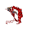
 Movie
Movie Controller


+ Open data

- Basic information
| Entry | Database: PDB / ID: 5b0g | ||||||
|---|---|---|---|---|---|---|---|
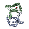
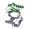
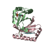
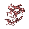
| Title | Polyketide cyclase OAC from Cannabis sativa, H78S mutant | ||||||
 Components Components | Olivetolic acid cyclase | ||||||
 Keywords Keywords | LYASE / Cannabis sativa / plant polyketide cyclase | ||||||
| Function / homology |  Function and homology informationolivetolic acid cyclase / olivetolic acid biosynthetic process / cannabinoid biosynthetic process / cyclase activity / terpenoid biosynthetic process / lyase activity / metal ion binding / cytoplasm Function and homology informationolivetolic acid cyclase / olivetolic acid biosynthetic process / cannabinoid biosynthetic process / cyclase activity / terpenoid biosynthetic process / lyase activity / metal ion binding / cytoplasmSimilarity search - Function | ||||||
| Biological species |  Cannabis sativa (plant) Cannabis sativa (plant) | ||||||
| Method | X-RAY DIFFRACTION / SYNCHROTRON / MOLECULAR REPLACEMENT / molecular replacement / Resolution: 1.4 Å | ||||||
 Authors Authors | Yang, X. / Matsui, T. / Mori, T. / Abe, I. / Morita, H. | ||||||
 Citation Citation | Journal: Febs J. / Year: 2016 Title: Structural basis for olivetolic acid formation by a polyketide cyclase from Cannabis sativa Authors: Yang, X. / Matsui, T. / Kodama, T. / Mori, T. / Zhou, X. / Taura, F. / Noguchi, H. / Abe, I. / Morita, H. | ||||||
| History |
|
- Structure visualization
| Structure viewer | Molecule: MolmilJmol/JSmol |
|---|
- Downloads & links
-Download
| PDBx/mmCIF format | 5b0g.cif.gz | 59.4 KB | Display | PDBx/mmCIF format |
|---|---|---|---|---|
| PDB format | pdb5b0g.ent.gz | 41.5 KB | Display | PDB format |
| PDBx/mmJSON format | 5b0g.json.gz | Tree view | PDBx/mmJSON format | |
| Others |  Other downloads Other downloads |
-Validation report
| Arichive directory | https://data.pdbj.org/pub/pdb/validation_reports/b0/5b0gftp://data.pdbj.org/pub/pdb/validation_reports/b0/5b0g | HTTPS FTP |
|---|
-Related structure data
| Related structure data |  5b08SC  5b09C  5b0aC  5b0bC  5b0cC  5b0dC  5b0eC  5b0fC C: citing same article ( S: Starting model for refinement |
|---|---|
| Similar structure data |


-Links
 PDBj
PDBj- Assembly
| Deposited unit | 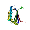
| ||||||||
|---|---|---|---|---|---|---|---|---|---|
| 1 | 
| ||||||||
| Unit cell |
| ||||||||
| Components on special symmetry positions |
|
-Components
| #1: Protein | Mass: 12178.941 Da / Num. of mol.: 1 / Mutation: H78S Source method: isolated from a genetically manipulated source Source: (gene. exp.) Cannabis sativa (plant) / Gene: OAC / Plasmid: pQE-80L / Production host:  Escherichia coli (E. coli) / Strain (production host): M15 / References: UniProt: I6WU39, olivetolic acid cyclase Escherichia coli (E. coli) / Strain (production host): M15 / References: UniProt: I6WU39, olivetolic acid cyclase |
|---|---|
| #2: Water | ChemComp-HOH / Water Mass: 18.015 Da / Num. of mol.: 155 / Source method: isolated from a natural source / Formula: H2O Mass: 18.015 Da / Num. of mol.: 155 / Source method: isolated from a natural source / Formula: H2O |
-Experimental details
-Experiment
| Experiment | Method: X-RAY DIFFRACTION / Number of used crystals: 1 |
|---|
- Sample preparation
| Crystal | Density Matthews: 2.01 Å3/Da / Density % sol: 38.81 % |
|---|---|
| Crystal grow | Temperature: 278 K / Method: vapor diffusion, sitting drop / Details: 100 mM MES pH 6.5, 30% (w/v) PEG4000 |
-Data collection
| Diffraction | Mean temperature: 100 K |
|---|---|
| Diffraction source | Source: SYNCHROTRON / Site: Photon Factory  / Beamline: AR-NW12A / Wavelength: 1 Å / Beamline: AR-NW12A / Wavelength: 1 Å |
| Detector | Type: ADSC QUANTUM 210r / Detector: CCD / Date: Dec 12, 2014 |
| Radiation | Monochromator: Si(1 1 1) / Protocol: SINGLE WAVELENGTH / Monochromatic (M) / Laue (L): M / Scattering type: x-ray |
| Radiation wavelength | Wavelength: 1 Å / Relative weight: 1 |
| Reflection | Resolution: 1.4→50 Å / Num. obs: 18918 / % possible obs: 99.2 % / Redundancy: 3.6 % / Rmerge(I) obs: 0.033 / Net I/σ(I): 25.3 |
| Reflection shell | Resolution: 1.4→1.48 Å / Redundancy: 3.5 % / Rmerge(I) obs: 0.13 / Mean I/σ(I) obs: 9.1 / % possible all: 98.7 |
-Phasing
| Phasing | Method: molecular replacement | ||||||
|---|---|---|---|---|---|---|---|
| Phasing MR | R rigid body: 0.594
|
- Processing
| Software |
| ||||||||||||||||||||||||||||||||||||||||||||||||||||||||||||||||||||||||||||||||||||||||||||||||||||||||||||||||||||||||||||||||||||||||||||||||||||||||||||||||||||||||||||||||||||||||||||||||||||||||
|---|---|---|---|---|---|---|---|---|---|---|---|---|---|---|---|---|---|---|---|---|---|---|---|---|---|---|---|---|---|---|---|---|---|---|---|---|---|---|---|---|---|---|---|---|---|---|---|---|---|---|---|---|---|---|---|---|---|---|---|---|---|---|---|---|---|---|---|---|---|---|---|---|---|---|---|---|---|---|---|---|---|---|---|---|---|---|---|---|---|---|---|---|---|---|---|---|---|---|---|---|---|---|---|---|---|---|---|---|---|---|---|---|---|---|---|---|---|---|---|---|---|---|---|---|---|---|---|---|---|---|---|---|---|---|---|---|---|---|---|---|---|---|---|---|---|---|---|---|---|---|---|---|---|---|---|---|---|---|---|---|---|---|---|---|---|---|---|---|---|---|---|---|---|---|---|---|---|---|---|---|---|---|---|---|---|---|---|---|---|---|---|---|---|---|---|---|---|---|---|---|---|
| Refinement | Method to determine structure: MOLECULAR REPLACEMENT Starting model: 5B08 Resolution: 1.4→40.362 Å / SU ML: 0.14 / Cross valid method: FREE R-VALUE / σ(F): 1.39 / Phase error: 19.61 / Stereochemistry target values: ML
| ||||||||||||||||||||||||||||||||||||||||||||||||||||||||||||||||||||||||||||||||||||||||||||||||||||||||||||||||||||||||||||||||||||||||||||||||||||||||||||||||||||||||||||||||||||||||||||||||||||||||
| Solvent computation | Shrinkage radii: 0.9 Å / VDW probe radii: 1.11 Å / Solvent model: FLAT BULK SOLVENT MODEL | ||||||||||||||||||||||||||||||||||||||||||||||||||||||||||||||||||||||||||||||||||||||||||||||||||||||||||||||||||||||||||||||||||||||||||||||||||||||||||||||||||||||||||||||||||||||||||||||||||||||||
| Displacement parameters | Biso max: 53.68 Å2 / Biso mean: 15.1684 Å2 / Biso min: 5.26 Å2 | ||||||||||||||||||||||||||||||||||||||||||||||||||||||||||||||||||||||||||||||||||||||||||||||||||||||||||||||||||||||||||||||||||||||||||||||||||||||||||||||||||||||||||||||||||||||||||||||||||||||||
| Refinement step | Cycle: final / Resolution: 1.4→40.362 Å
| ||||||||||||||||||||||||||||||||||||||||||||||||||||||||||||||||||||||||||||||||||||||||||||||||||||||||||||||||||||||||||||||||||||||||||||||||||||||||||||||||||||||||||||||||||||||||||||||||||||||||
| Refine LS restraints |
| ||||||||||||||||||||||||||||||||||||||||||||||||||||||||||||||||||||||||||||||||||||||||||||||||||||||||||||||||||||||||||||||||||||||||||||||||||||||||||||||||||||||||||||||||||||||||||||||||||||||||
| LS refinement shell | Refine-ID: X-RAY DIFFRACTION / Total num. of bins used: 7
| ||||||||||||||||||||||||||||||||||||||||||||||||||||||||||||||||||||||||||||||||||||||||||||||||||||||||||||||||||||||||||||||||||||||||||||||||||||||||||||||||||||||||||||||||||||||||||||||||||||||||
| Refinement TLS params. | Method: refined / Refine-ID: X-RAY DIFFRACTION
| ||||||||||||||||||||||||||||||||||||||||||||||||||||||||||||||||||||||||||||||||||||||||||||||||||||||||||||||||||||||||||||||||||||||||||||||||||||||||||||||||||||||||||||||||||||||||||||||||||||||||
| Refinement TLS group |
|
