 Movie
Movie Controller

[English] 日本語
 Yorodumi
- PDB-5az7: Crystal structure of MBP-Tom20 fusion protein with a 4-residue sp... -
+ Open data

- Basic information
| Entry | Database: PDB / ID: 5az7 | |||||||||
|---|---|---|---|---|---|---|---|---|---|---|
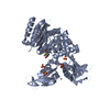
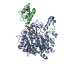
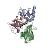
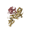
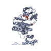
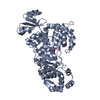
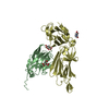
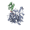
| Title | Crystal structure of MBP-Tom20 fusion protein with a 4-residue spacer in the connector helix | |||||||||
 Components Components | Maltose-binding periplasmic protein,Mitochondrial import receptor subunit TOM20 homolog | |||||||||
 Keywords Keywords | SUGAR BINDING PROTEIN / PEPTIDE BINDING PROTEIN /  Fusion protein Fusion protein | |||||||||
| Function / homology |  Function and homology information Function and homology informationtRNA import into mitochondrion / PINK1-PRKN Mediated Mitophagy / mitochondrion targeting sequence binding / mitochondrial outer membrane translocase complex / response to 3,3',5-triiodo-L-thyronine / mitochondria-associated endoplasmic reticulum membrane contact site / migrasome / Ub-specific processing proteases / protein import into mitochondrial matrix / protein-transporting ATPase activity ...tRNA import into mitochondrion / PINK1-PRKN Mediated Mitophagy / mitochondrion targeting sequence binding / mitochondrial outer membrane translocase complex / response to 3,3',5-triiodo-L-thyronine / mitochondria-associated endoplasmic reticulum membrane contact site / migrasome / Ub-specific processing proteases / protein import into mitochondrial matrix / protein-transporting ATPase activity / protein targeting to mitochondrion / mitochondrial envelope / response to muscle activity / detection of maltose stimulus / maltose transport complex / maltose binding / maltose transport / maltodextrin transmembrane transport / carbohydrate transport / carbohydrate transmembrane transporter activity / ATP-binding cassette (ABC) transporter complex, substrate-binding subunit-containing / sperm midpiece / ATP-binding cassette (ABC) transporter complex / cell chemotaxis / cell periphery / intracellular protein transport / unfolded protein binding / outer membrane-bounded periplasmic space / mitochondrial outer membrane / periplasmic space / DNA damage response / mitochondrion / membraneSimilarity search - Function | |||||||||
| Biological species |  Escherichia coli (E. coli) Escherichia coli (E. coli) Rattus norvegicus (Norway rat) Rattus norvegicus (Norway rat) | |||||||||
| Method | X-RAY DIFFRACTION / SYNCHROTRON / MOLECULAR REPLACEMENT / Resolution: 1.96 Å | |||||||||
 Authors Authors | Matsuoka, R. / Kohda, D. | |||||||||
 Citation Citation | Journal: Protein Sci. / Year: 2016 Title: Rational design of crystal contact-free space in protein crystals for analyzing spatial distribution of motions within protein molecules. Authors: Matsuoka, R. / Shimada, A. / Komuro, Y. / Sugita, Y. / Kohda, D. | |||||||||
| History |
|
- Structure visualization
| Structure viewer | Molecule: MolmilJmol/JSmol |
|---|
- Downloads & links
-Download
| PDBx/mmCIF format | 5az7.cif.gz | 101 KB | Display | PDBx/mmCIF format |
|---|---|---|---|---|
| PDB format | pdb5az7.ent.gz | 75 KB | Display | PDB format |
| PDBx/mmJSON format | 5az7.json.gz | Tree view | PDBx/mmJSON format | |
| Others |  Other downloads Other downloads |
-Validation report
| Arichive directory | https://data.pdbj.org/pub/pdb/validation_reports/az/5az7ftp://data.pdbj.org/pub/pdb/validation_reports/az/5az7 | HTTPS FTP |
|---|
-Related structure data
| Related structure data | 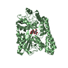 5az6C  5az8C  5az9C 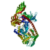 5azaC  1anfS  2v1tS C: citing same article ( S: Starting model for refinement |
|---|---|
| Similar structure data |


-Links
 PDBj
PDBj


- Assembly
| Deposited unit | 
| ||||||||
|---|---|---|---|---|---|---|---|---|---|
| 1 |
| ||||||||
| Unit cell |
| ||||||||
| Components on special symmetry positions |
|
-Components
| #1: Protein | Mass: 48028.551 Da / Num. of mol.: 1 / Fragment: UNP RESIDUES 27-394,UNP RESIDUES 65-126 / Mutation: A313V, C409S,A313V, C409S Source method: isolated from a genetically manipulated source Details: The fusion protein of 1-369 Maltose binding protein, 370-373 Linker and 374-435 Tom20 Source: (gene. exp.) Escherichia coli (strain K12) (bacteria), (gene. exp.) Rattus norvegicus (Norway rat)Strain: K12 / Gene: malE, b4034, JW3994, Tomm20 / Production host: Escherichia coli (E. coli) / References: UniProt: P0AEX9, UniProt: Q62760 |
|---|---|
| #2: Polysaccharide | alpha-D-glucopyranose-(1-4)-alpha-D-glucopyranose / alpha-maltose  , Oligosaccharide / Class: Nutrient / Mass: 342.297 Da / Num. of mol.: 1 , Oligosaccharide / Class: Nutrient / Mass: 342.297 Da / Num. of mol.: 1Source method: isolated from a genetically manipulated source Details: oligosaccharide / References: alpha-maltose |
| #3: Water | ChemComp-HOH / Water Mass: 18.015 Da / Num. of mol.: 101 / Source method: isolated from a natural source / Formula: H2O Mass: 18.015 Da / Num. of mol.: 101 / Source method: isolated from a natural source / Formula: H2O |
-Experimental details
-Experiment
| Experiment | Method: X-RAY DIFFRACTION |
|---|
- Sample preparation
| Crystal | Density Matthews: 2.35 Å3/Da / Density % sol: 47.73 % |
|---|---|
| Crystal grow | Temperature: 293 K / Method: vapor diffusion, hanging drop / pH: 4.4 / Details: 50% PEG 400, 0.1M Phosphate citrate (pH 4.4) |
-Data collection
| Diffraction | Mean temperature: 100 K |
|---|---|
| Diffraction source | Source: SYNCHROTRON / Site: Photon Factory  / Beamline: BL-5A / Wavelength: 1 Å / Beamline: BL-5A / Wavelength: 1 Å |
| Detector | Type: ADSC QUANTUM 210 / Detector: CCD / Date: Jan 24, 2012 |
| Radiation | Protocol: SINGLE WAVELENGTH / Monochromatic (M) / Laue (L): M / Scattering type: x-ray |
| Radiation wavelength | Wavelength: 1 Å / Relative weight: 1 |
| Reflection | Resolution: 1.96→50 Å / Num. obs: 30663 / % possible obs: 93.1 % / Redundancy: 6.14 % / Net I/σ(I): 14.6 |
- Processing
| Software |
| ||||||||||||||||||||||||||||||||||||||||||||||||||||||||||||||||||||||||||||||||||||||||||||||||||||||||||||||||||||||||||||||||||||||||||||||||||||||||||||||||||||||||||||||||||||||
|---|---|---|---|---|---|---|---|---|---|---|---|---|---|---|---|---|---|---|---|---|---|---|---|---|---|---|---|---|---|---|---|---|---|---|---|---|---|---|---|---|---|---|---|---|---|---|---|---|---|---|---|---|---|---|---|---|---|---|---|---|---|---|---|---|---|---|---|---|---|---|---|---|---|---|---|---|---|---|---|---|---|---|---|---|---|---|---|---|---|---|---|---|---|---|---|---|---|---|---|---|---|---|---|---|---|---|---|---|---|---|---|---|---|---|---|---|---|---|---|---|---|---|---|---|---|---|---|---|---|---|---|---|---|---|---|---|---|---|---|---|---|---|---|---|---|---|---|---|---|---|---|---|---|---|---|---|---|---|---|---|---|---|---|---|---|---|---|---|---|---|---|---|---|---|---|---|---|---|---|---|---|---|---|
| Refinement | Method to determine structure: MOLECULAR REPLACEMENT Starting model: 1ANF, 2V1T Resolution: 1.96→42.22 Å / Cor.coef. Fo:Fc: 0.938 / Cor.coef. Fo:Fc free: 0.921 / SU B: 4.497 / SU ML: 0.124 / Cross valid method: THROUGHOUT / ESU R: 0.193 / ESU R Free: 0.172 / Stereochemistry target values: MAXIMUM LIKELIHOOD Details: HYDROGENS HAVE BEEN ADDED IN THE RIDING POSITIONS U VALUES
| ||||||||||||||||||||||||||||||||||||||||||||||||||||||||||||||||||||||||||||||||||||||||||||||||||||||||||||||||||||||||||||||||||||||||||||||||||||||||||||||||||||||||||||||||||||||
| Solvent computation | Ion probe radii: 0.8 Å / Shrinkage radii: 0.8 Å / VDW probe radii: 1.2 Å / Solvent model: MASK | ||||||||||||||||||||||||||||||||||||||||||||||||||||||||||||||||||||||||||||||||||||||||||||||||||||||||||||||||||||||||||||||||||||||||||||||||||||||||||||||||||||||||||||||||||||||
| Displacement parameters | Biso mean: 35.961 Å2
| ||||||||||||||||||||||||||||||||||||||||||||||||||||||||||||||||||||||||||||||||||||||||||||||||||||||||||||||||||||||||||||||||||||||||||||||||||||||||||||||||||||||||||||||||||||||
| Refinement step | Cycle: LAST / Resolution: 1.96→42.22 Å
| ||||||||||||||||||||||||||||||||||||||||||||||||||||||||||||||||||||||||||||||||||||||||||||||||||||||||||||||||||||||||||||||||||||||||||||||||||||||||||||||||||||||||||||||||||||||
| Refine LS restraints |
|