 Movie
Movie Controller

[English] 日本語
 Yorodumi
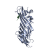
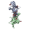
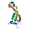
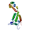
- PDB-5awt: Crystal structure of the SGIP1 mu homology domain in complex with... -
+ Open data

- Basic information
| Entry | Database: PDB / ID: 5awt | ||||||||||||
|---|---|---|---|---|---|---|---|---|---|---|---|---|---|
| Title | Crystal structure of the SGIP1 mu homology domain in complex with an Eps15 fragment containing two DPF motifs (YDPFGGDPFKG) | ||||||||||||
 Components Components |
| ||||||||||||
 Keywords Keywords |  ENDOCYTOSIS / Protein-protein interaction ENDOCYTOSIS / Protein-protein interaction | ||||||||||||
| Function / homology |  Function and homology information Function and homology informationpositive regulation of feeding behavior / Golgi to endosome transport / AP-2 adaptor complex / clathrin coat of coated pit / vesicle organization / postsynaptic neurotransmitter receptor internalization / clathrin coat assembly / clathrin-dependent endocytosis / endocytic recycling / aggresome ...positive regulation of feeding behavior / Golgi to endosome transport / AP-2 adaptor complex / clathrin coat of coated pit / vesicle organization / postsynaptic neurotransmitter receptor internalization / clathrin coat assembly / clathrin-dependent endocytosis / endocytic recycling / aggresome / clathrin-coated vesicle / endosomal transport / positive regulation of receptor recycling / response to dietary excess / polyubiquitin modification-dependent protein binding / energy homeostasis / clathrin-coated pit / basal plasma membrane / InlB-mediated entry of Listeria monocytogenes into host cell / EGFR downregulation / Negative regulation of MET activity / phospholipid binding / SH3 domain binding / positive regulation of receptor-mediated endocytosis / endocytosis / protein transport / Cargo recognition for clathrin-mediated endocytosis / Clathrin-mediated endocytosis / regulation of cell population proliferation / early endosome membrane / postsynapse / microtubule binding / receptor-mediated endocytosis of virus by host cell / symbiont entry into host cell / cadherin binding / apical plasma membrane / intracellular membrane-bounded organelle / glutamatergic synapse / calcium ion binding / membrane / plasma membrane / cytosol / cytoplasmSimilarity search - Function | ||||||||||||
| Biological species |  Homo sapiens (human) Homo sapiens (human) | ||||||||||||
| Method | X-RAY DIFFRACTION / SYNCHROTRON / MOLECULAR REPLACEMENT / Resolution: 2.702 Å | ||||||||||||
 Authors Authors | Shimada, A. / Yamaguchi, A. / Kohda, D. | ||||||||||||
| Funding support |  Japan, 3items Japan, 3items
| ||||||||||||
 Citation Citation | Journal: Sci Rep / Year: 2016 Title: Structural basis for the recognition of two consecutive mutually interacting DPF motifs by the SGIP1 mu homology domain. Authors: Shimada, A. / Yamaguchi, A. / Kohda, D. | ||||||||||||
| History |
|
- Structure visualization
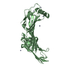
| Structure viewer | Molecule: MolmilJmol/JSmol |
|---|
- Downloads & links
-Download
| PDBx/mmCIF format | 5awt.cif.gz | 128.1 KB | Display | PDBx/mmCIF format |
|---|---|---|---|---|
| PDB format | pdb5awt.ent.gz | 98.6 KB | Display | PDB format |
| PDBx/mmJSON format | 5awt.json.gz | Tree view | PDBx/mmJSON format | |
| Others |  Other downloads Other downloads |
-Validation report
| Arichive directory | https://data.pdbj.org/pub/pdb/validation_reports/aw/5awtftp://data.pdbj.org/pub/pdb/validation_reports/aw/5awt | HTTPS FTP |
|---|
-Related structure data
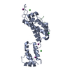
| Related structure data |  5awrSC  5awsC 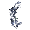 5awuC S: Starting model for refinement C: citing same article ( |
|---|---|
| Similar structure data |


-Links
 PDBj
PDBj


- Assembly
| Deposited unit | 
| ||||||||
|---|---|---|---|---|---|---|---|---|---|
| 1 |
| ||||||||
| Unit cell |
|
-Components
| #1: Protein | Mass: 30844.158 Da / Num. of mol.: 1 / Fragment: UNP RESIDUES 552-828 Source method: isolated from a genetically manipulated source Source: (gene. exp.) Homo sapiens (human) / Gene: SGIP1 / Plasmid: pGEX-6P-1 / Production host:  Escherichia coli (E. coli) / References: UniProt: Q9BQI5 Escherichia coli (E. coli) / References: UniProt: Q9BQI5 | ||
|---|---|---|---|
| #2: Protein/peptide | Mass: 1200.277 Da / Num. of mol.: 1 / Source method: obtained synthetically / Source: (synth.) Homo sapiens (human) / References: UniProt: P42566*PLUS | ||
| #3: Chemical | ChemComp-ZN /   Mass: 65.409 Da / Num. of mol.: 6 / Source method: obtained synthetically / Formula: Zn Mass: 65.409 Da / Num. of mol.: 6 / Source method: obtained synthetically / Formula: Zn#4: Water | ChemComp-HOH / | Water Mass: 18.015 Da / Num. of mol.: 35 / Source method: isolated from a natural source / Formula: H2O Mass: 18.015 Da / Num. of mol.: 35 / Source method: isolated from a natural source / Formula: H2O |
-Experimental details
-Experiment
| Experiment | Method: X-RAY DIFFRACTION |
|---|
- Sample preparation
| Crystal | Density Matthews: 3.62 Å3/Da / Density % sol: 66.02 % |
|---|---|
| Crystal grow | Temperature: 293 K / Method: vapor diffusion, hanging drop / pH: 5.1 Details: PEG 3350, zinc acetate, sodium acetate, sodium iodide |
-Data collection
| Diffraction | Mean temperature: 100 K |
|---|---|
| Diffraction source | Source: SYNCHROTRON / Site: SPring-8 / Beamline: BL26B2 / Wavelength: 1 Å |
| Detector | Type: RAYONIX MX225HE / Detector: CCD / Date: Dec 17, 2013 |
| Radiation | Protocol: SINGLE WAVELENGTH / Monochromatic (M) / Laue (L): M / Scattering type: x-ray |
| Radiation wavelength | Wavelength: 1 Å / Relative weight: 1 |
| Reflection | Resolution: 2.7→50 Å / Num. obs: 13428 / % possible obs: 100 % / Redundancy: 14.1 % / Net I/σ(I): 26.8 |
- Processing
| Software |
| ||||||||||||||||||||||||||||||||||||||||||||||||||||||||||||||||||||||||||||||||||||||||||||||||||||||||||||||||||||||||||||||||||||||||||||||||||||||||||||||||||||||||||||||||||||||||||||||||||||||||
|---|---|---|---|---|---|---|---|---|---|---|---|---|---|---|---|---|---|---|---|---|---|---|---|---|---|---|---|---|---|---|---|---|---|---|---|---|---|---|---|---|---|---|---|---|---|---|---|---|---|---|---|---|---|---|---|---|---|---|---|---|---|---|---|---|---|---|---|---|---|---|---|---|---|---|---|---|---|---|---|---|---|---|---|---|---|---|---|---|---|---|---|---|---|---|---|---|---|---|---|---|---|---|---|---|---|---|---|---|---|---|---|---|---|---|---|---|---|---|---|---|---|---|---|---|---|---|---|---|---|---|---|---|---|---|---|---|---|---|---|---|---|---|---|---|---|---|---|---|---|---|---|---|---|---|---|---|---|---|---|---|---|---|---|---|---|---|---|---|---|---|---|---|---|---|---|---|---|---|---|---|---|---|---|---|---|---|---|---|---|---|---|---|---|---|---|---|---|---|---|---|---|
| Refinement | Method to determine structure: MOLECULAR REPLACEMENT Starting model: 5AWR Resolution: 2.702→41.265 Å / SU ML: 0.32 / Cross valid method: FREE R-VALUE / σ(F): 1.36 / Phase error: 23.04 / Stereochemistry target values: ML
| ||||||||||||||||||||||||||||||||||||||||||||||||||||||||||||||||||||||||||||||||||||||||||||||||||||||||||||||||||||||||||||||||||||||||||||||||||||||||||||||||||||||||||||||||||||||||||||||||||||||||
| Solvent computation | Shrinkage radii: 0.9 Å / VDW probe radii: 1.11 Å / Solvent model: FLAT BULK SOLVENT MODEL | ||||||||||||||||||||||||||||||||||||||||||||||||||||||||||||||||||||||||||||||||||||||||||||||||||||||||||||||||||||||||||||||||||||||||||||||||||||||||||||||||||||||||||||||||||||||||||||||||||||||||
| Refinement step | Cycle: LAST / Resolution: 2.702→41.265 Å
| ||||||||||||||||||||||||||||||||||||||||||||||||||||||||||||||||||||||||||||||||||||||||||||||||||||||||||||||||||||||||||||||||||||||||||||||||||||||||||||||||||||||||||||||||||||||||||||||||||||||||
| Refine LS restraints |
| ||||||||||||||||||||||||||||||||||||||||||||||||||||||||||||||||||||||||||||||||||||||||||||||||||||||||||||||||||||||||||||||||||||||||||||||||||||||||||||||||||||||||||||||||||||||||||||||||||||||||
| LS refinement shell |
| ||||||||||||||||||||||||||||||||||||||||||||||||||||||||||||||||||||||||||||||||||||||||||||||||||||||||||||||||||||||||||||||||||||||||||||||||||||||||||||||||||||||||||||||||||||||||||||||||||||||||
| Refinement TLS params. | Method: refined / Refine-ID: X-RAY DIFFRACTION
| ||||||||||||||||||||||||||||||||||||||||||||||||||||||||||||||||||||||||||||||||||||||||||||||||||||||||||||||||||||||||||||||||||||||||||||||||||||||||||||||||||||||||||||||||||||||||||||||||||||||||
| Refinement TLS group |
|
