| Entry | Database: PDB / ID: 5ava
|
|---|
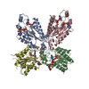
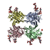
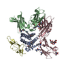
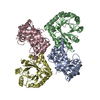
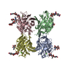
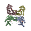
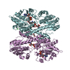
| Title | Crystal structure of PHA-E lectin in complex with bisected glycan |
|---|
 Components Components | Erythroagglutinin |
|---|
 Keywords Keywords | SUGAR BINDING PROTEIN /  lectin / glycan lectin / glycan |
|---|
| Function / homology |  Function and homology information Function and homology information |
|---|
| Biological species |  Phaseolus vulgaris (French bean) Phaseolus vulgaris (French bean) |
|---|
| Method | X-RAY DIFFRACTION / SYNCHROTRON / MOLECULAR REPLACEMENT / Resolution: 3 Å |
|---|
 Authors Authors | Nagae, M. / Yamaguchi, Y. |
|---|
| Funding support |  Japan, 1items Japan, 1items | Organization | Grant number | Country |
|---|
| MEXT | 15K18496, 26110724 | Japan |
|
|---|
 Citation Citation | Journal: Sci Rep / Year: 2016
Title: Atomic visualization of a flipped-back conformation of bisected glycans bound to specific lectins
Authors: Nagae, M. / Kanagawa, M. / Morita-Matsumoto, K. / Hanashima, S. / Kizuka, Y. / Taniguchi, N. / Yamaguchi, Y. |
|---|
| History | | Deposition | Jun 12, 2015 | Deposition site: PDBJ / Processing site: PDBJ |
|---|
| Revision 1.0 | Apr 27, 2016 | Provider: repository / Type: Initial release |
|---|
| Revision 1.1 | Feb 19, 2020 | Group: Data collection / Derived calculations / Category: chem_comp / diffrn_source / pdbx_struct_oper_list
Item: _chem_comp.type / _diffrn_source.pdbx_synchrotron_site / _pdbx_struct_oper_list.symmetry_operation |
|---|
| Revision 2.0 | Jul 29, 2020 | Group: Advisory / Atomic model ...Advisory / Atomic model / Data collection / Derived calculations / Structure summary
Category: atom_site / chem_comp ...atom_site / chem_comp / entity / pdbx_branch_scheme / pdbx_chem_comp_identifier / pdbx_entity_branch / pdbx_entity_branch_descriptor / pdbx_entity_branch_link / pdbx_entity_branch_list / pdbx_entity_nonpoly / pdbx_nonpoly_scheme / pdbx_struct_assembly_gen / pdbx_struct_conn_angle / pdbx_struct_mod_residue / pdbx_unobs_or_zero_occ_atoms / struct_asym / struct_conn / struct_conn_type / struct_site / struct_site_gen
Item: _atom_site.B_iso_or_equiv / _atom_site.Cartn_x ..._atom_site.B_iso_or_equiv / _atom_site.Cartn_x / _atom_site.Cartn_y / _atom_site.Cartn_z / _atom_site.auth_asym_id / _atom_site.auth_atom_id / _atom_site.auth_comp_id / _atom_site.auth_seq_id / _atom_site.label_asym_id / _atom_site.label_atom_id / _atom_site.label_comp_id / _atom_site.label_entity_id / _atom_site.type_symbol / _chem_comp.name / _pdbx_struct_assembly_gen.asym_id_list / _pdbx_struct_conn_angle.ptnr1_auth_seq_id / _pdbx_struct_conn_angle.ptnr1_label_asym_id / _pdbx_struct_conn_angle.ptnr2_label_asym_id / _pdbx_struct_conn_angle.ptnr3_auth_seq_id / _pdbx_struct_conn_angle.ptnr3_label_asym_id / _pdbx_struct_conn_angle.value / _pdbx_struct_mod_residue.auth_asym_id / _pdbx_struct_mod_residue.auth_seq_id / _pdbx_struct_mod_residue.label_asym_id / _pdbx_unobs_or_zero_occ_atoms.auth_asym_id / _pdbx_unobs_or_zero_occ_atoms.auth_seq_id / _pdbx_unobs_or_zero_occ_atoms.label_asym_id / _struct_conn.conn_type_id / _struct_conn.id / _struct_conn.pdbx_dist_value / _struct_conn.pdbx_leaving_atom_flag / _struct_conn.ptnr1_auth_asym_id / _struct_conn.ptnr1_auth_comp_id / _struct_conn.ptnr1_auth_seq_id / _struct_conn.ptnr1_label_asym_id / _struct_conn.ptnr1_label_atom_id / _struct_conn.ptnr1_label_comp_id / _struct_conn.ptnr1_label_seq_id / _struct_conn.ptnr2_auth_asym_id / _struct_conn.ptnr2_auth_comp_id / _struct_conn.ptnr2_auth_seq_id / _struct_conn.ptnr2_label_asym_id / _struct_conn.ptnr2_label_atom_id / _struct_conn.ptnr2_label_comp_id / _struct_conn_type.id
Description: Carbohydrate remediation / Provider: repository / Type: Remediation |
|---|
| Revision 2.1 | Nov 8, 2023 | Group: Data collection / Database references ...Data collection / Database references / Derived calculations / Refinement description / Structure summary
Category: chem_comp / chem_comp_atom ...chem_comp / chem_comp_atom / chem_comp_bond / database_2 / pdbx_initial_refinement_model / struct_conn
Item: _chem_comp.pdbx_synonyms / _database_2.pdbx_DOI ..._chem_comp.pdbx_synonyms / _database_2.pdbx_DOI / _database_2.pdbx_database_accession / _struct_conn.pdbx_leaving_atom_flag |
|---|
|
|---|
 Movie
Movie Controller

 Yorodumi
 Open data
 Basic information
 Structure visualization
 Downloads & links
 Other downloads
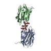


 PDBj
PDBj

 Assembly
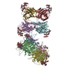


 Mass: 54.938 Da / Num. of mol.: 8 / Source method: obtained synthetically / Formula: Mn
 Mass: 40.078 Da / Num. of mol.: 8 / Source method: obtained synthetically / Formula: Ca
 Mass: 18.015 Da / Num. of mol.: 32 / Source method: isolated from a natural source / Formula: H2O
 Sample preparation
 Processing