 Movie
Movie Controller

[English] 日本語
 Yorodumi
- PDB-5aq8: DARPin-based Crystallization Chaperones exploit Molecular Geometr... -
+ Open data

- Basic information
| Entry | Database: PDB / ID: 5aq8 | ||||||
|---|---|---|---|---|---|---|---|
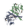
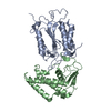
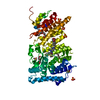
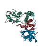
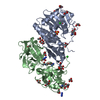
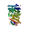
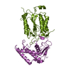
| Title | DARPin-based Crystallization Chaperones exploit Molecular Geometry as a Screening Dimension in Protein Crystallography | ||||||
 Components Components | OFF7_DB12V4 | ||||||
 Keywords Keywords |  CHAPERONE / CRYSTALLIZATION CHAPERONE / DESIGNED ANKYRIN REPEAT PROTEIN (DARPIN) / RIGID DOMAIN FUSION CHAPERONE / CRYSTALLIZATION CHAPERONE / DESIGNED ANKYRIN REPEAT PROTEIN (DARPIN) / RIGID DOMAIN FUSION | ||||||
| Function / homology | Ankyrin repeat-containing domain / Beta-lactamase / DD-peptidase/beta-lactamase superfamily / Serine Threonine Protein Phosphatase 5, Tetratricopeptide repeat / Alpha Horseshoe / 3-Layer(aba) Sandwich / Mainly Alpha / Alpha Beta / THIOCYANATE ION Function and homology information Function and homology information | ||||||
| Biological species | SYNTHETIC CONSTRUCT (others) | ||||||
| Method | X-RAY DIFFRACTION / SYNCHROTRON / MOLECULAR REPLACEMENT / Resolution: 1.62 Å | ||||||
 Authors Authors | Batyuk, A. / Wu, Y. / Honegger, A. / Heberling, M. / Plueckthun, A. | ||||||
 Citation Citation | Journal: J.Mol.Biol. / Year: 2016 Title: Darpin-Based Crystallization Chaperones Exploit Molecular Geometry as a Screening Dimension in Protein Crystallography Authors: Batyuk, A. / Wu, Y. / Honegger, A. / Heberling, M. / Plueckthun, A. | ||||||
| History |
|
- Structure visualization
| Structure viewer | Molecule: MolmilJmol/JSmol |
|---|
- Downloads & links
-Download
| PDBx/mmCIF format | 5aq8.cif.gz | 240.9 KB | Display | PDBx/mmCIF format |
|---|---|---|---|---|
| PDB format | pdb5aq8.ent.gz | 198.7 KB | Display | PDB format |
| PDBx/mmJSON format | 5aq8.json.gz | Tree view | PDBx/mmJSON format | |
| Others |  Other downloads Other downloads |
-Validation report
| Arichive directory | https://data.pdbj.org/pub/pdb/validation_reports/aq/5aq8ftp://data.pdbj.org/pub/pdb/validation_reports/aq/5aq8 | HTTPS FTP |
|---|
-Related structure data
| Related structure data |  5aq7C  5aq9C  5aqaC  5aqbC  1svxS 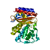 3dtmS C: citing same article ( S: Starting model for refinement |
|---|---|
| Similar structure data |

-Links
 PDBj
PDBj

- Assembly
| Deposited unit | 
| ||||||||
|---|---|---|---|---|---|---|---|---|---|
| 1 |
| ||||||||
| Unit cell |
|
-Components
| #1: Protein | Mass: 45962.926 Da / Num. of mol.: 1 Source method: isolated from a genetically manipulated source Source: (gene. exp.) SYNTHETIC CONSTRUCT (others) / Plasmid: PQE30 / Production host:  ESCHERICHIA COLI (E. coli) / Strain (production host): K-12 / Variant (production host): XL1-BLUE ESCHERICHIA COLI (E. coli) / Strain (production host): K-12 / Variant (production host): XL1-BLUE | ||||
|---|---|---|---|---|---|
| #2: Chemical | HEPES  Mass: 238.305 Da / Num. of mol.: 2 / Source method: obtained synthetically / Formula: C8H18N2O4S / Comment: pH buffer*YM Mass: 238.305 Da / Num. of mol.: 2 / Source method: obtained synthetically / Formula: C8H18N2O4S / Comment: pH buffer*YM#3: Chemical | Thiocyanate  Mass: 58.082 Da / Num. of mol.: 3 / Source method: obtained synthetically / Formula: CNS Mass: 58.082 Da / Num. of mol.: 3 / Source method: obtained synthetically / Formula: CNS#4: Water | ChemComp-HOH / | Water Mass: 18.015 Da / Num. of mol.: 289 / Source method: isolated from a natural source / Formula: H2O Mass: 18.015 Da / Num. of mol.: 289 / Source method: isolated from a natural source / Formula: H2O |
-Experimental details
-Experiment
| Experiment | Method: X-RAY DIFFRACTION / Number of used crystals: 1 |
|---|
- Sample preparation
| Crystal | Density Matthews: 1.75 Å3/Da / Density % sol: 29.7 % / Description: NONE |
|---|---|
| Crystal grow | pH: 7.5 Details: PEG4000 18.3 %, KSCN 0.286 M, HEPES-NA, 0.1 M, PH 7.5 |
-Data collection
| Diffraction | Mean temperature: 100 K |
|---|---|
| Diffraction source | Source: SYNCHROTRON / Site: SLS  / Beamline: X06SA / Wavelength: 1 / Beamline: X06SA / Wavelength: 1 |
| Detector | Type: DECTRIS PILATUS 6M / Detector: PIXEL / Date: Nov 24, 2011 / Details: RH COATED MERIDIONALLY FOCUSSING MIRROR |
| Radiation | Monochromator: FIXED-EXIT LN2 COOLED DOUBLE CRYSTAL MONOCHROMATOR Protocol: SINGLE WAVELENGTH / Monochromatic (M) / Laue (L): M / Scattering type: x-ray |
| Radiation wavelength | Wavelength: 1 Å / Relative weight: 1 |
| Reflection | Resolution: 1.62→43.72 Å / Num. obs: 43423 / % possible obs: 1 % / Observed criterion σ(I): 2 / Redundancy: 6.3 % / Biso Wilson estimate: 18.64 Å2 / Rmerge(I) obs: 0.12 / Net I/σ(I): 11.13 |
| Reflection shell | Resolution: 1.62→1.68 Å / Redundancy: 6 % / Mean I/σ(I) obs: 1.51 / % possible all: 99.4 |
- Processing
| Software |
| |||||||||||||||||||||||||||||||||||||||||||||||||||||||||||||||||||||||||||||||||||||||||||||||||||||||||||||||||||||||
|---|---|---|---|---|---|---|---|---|---|---|---|---|---|---|---|---|---|---|---|---|---|---|---|---|---|---|---|---|---|---|---|---|---|---|---|---|---|---|---|---|---|---|---|---|---|---|---|---|---|---|---|---|---|---|---|---|---|---|---|---|---|---|---|---|---|---|---|---|---|---|---|---|---|---|---|---|---|---|---|---|---|---|---|---|---|---|---|---|---|---|---|---|---|---|---|---|---|---|---|---|---|---|---|---|---|---|---|---|---|---|---|---|---|---|---|---|---|---|---|---|
| Refinement | Method to determine structure: MOLECULAR REPLACEMENT Starting model: PDB ENTRIES 3DTM AND 1SVX CHAIN A Resolution: 1.62→43.717 Å / SU ML: 0.21 / σ(F): 1.36 / Phase error: 26.86 / Stereochemistry target values: ML
| |||||||||||||||||||||||||||||||||||||||||||||||||||||||||||||||||||||||||||||||||||||||||||||||||||||||||||||||||||||||
| Solvent computation | Shrinkage radii: 0.9 Å / VDW probe radii: 1.11 Å / Solvent model: FLAT BULK SOLVENT MODEL | |||||||||||||||||||||||||||||||||||||||||||||||||||||||||||||||||||||||||||||||||||||||||||||||||||||||||||||||||||||||
| Displacement parameters | Biso mean: 26.04 Å2 | |||||||||||||||||||||||||||||||||||||||||||||||||||||||||||||||||||||||||||||||||||||||||||||||||||||||||||||||||||||||
| Refinement step | Cycle: LAST / Resolution: 1.62→43.717 Å
| |||||||||||||||||||||||||||||||||||||||||||||||||||||||||||||||||||||||||||||||||||||||||||||||||||||||||||||||||||||||
| Refine LS restraints |
| |||||||||||||||||||||||||||||||||||||||||||||||||||||||||||||||||||||||||||||||||||||||||||||||||||||||||||||||||||||||
| LS refinement shell |
|
