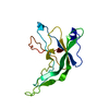
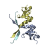
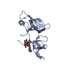
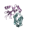
[English] 日本語
Yorodumi
- PDB-5aq0: The structure of the Transthyretin-like domain of the first catal... -

+
Open data


ID or keywords:

Loading...

-
Basic information

Entry
Database: PDB / ID: 5aq0
TitleThe structure of the Transthyretin-like domain of the first catalytic domain of the HUMAN Carboxypeptidase D
ComponentsCARBOXYPEPTIDASE D
KeywordsHYDROLASE / MAN CARBOXYPEPTIDASE D / TTL / TRANSTHYRETIN-LIKE DOMAIN
Function / homology
Function and homology information


metallocarboxypeptidase D / serine-type carboxypeptidase activity / peptide metabolic process / Golgi Associated Vesicle Biogenesis / metallocarboxypeptidase activity / protein processing / membrane => GO:0016020 / extracellular space / extracellular exosome / zinc ion binding ...metallocarboxypeptidase D / serine-type carboxypeptidase activity / peptide metabolic process / Golgi Associated Vesicle Biogenesis / metallocarboxypeptidase activity / protein processing / membrane => GO:0016020 / extracellular space / extracellular exosome / zinc ion binding / membrane / plasma membrane
Similarity search - Function
Peptidase M14B, caboxypeptidase D / Carboxypeptidase D, carboxypeptidase-like domain 2 / Carboxypeptidase regulatory-like domain / Carboxypeptidase-like, regulatory domain / Carboxypeptidase-like, regulatory domain superfamily / Zinc carboxypeptidases, zinc-binding region 2 signature. / Zinc carboxypeptidases, zinc-binding region 1 signature. / Zn_pept / Peptidase M14, carboxypeptidase A / Zinc carboxypeptidase ...Peptidase M14B, caboxypeptidase D / Carboxypeptidase D, carboxypeptidase-like domain 2 / Carboxypeptidase regulatory-like domain / Carboxypeptidase-like, regulatory domain / Carboxypeptidase-like, regulatory domain superfamily / Zinc carboxypeptidases, zinc-binding region 2 signature. / Zinc carboxypeptidases, zinc-binding region 1 signature. / Zn_pept / Peptidase M14, carboxypeptidase A / Zinc carboxypeptidase / Immunoglobulin-like / Sandwich / Mainly Beta
Similarity search - Domain/homology
Biological speciesHOMO SAPIENS (human)
MethodX-RAY DIFFRACTION / SYNCHROTRON / MOLECULAR REPLACEMENT / Resolution: 0.95 Å
AuthorsGallego, P. / Garcia-Pardo, J. / Lorenzo, J. / Aviles, F.X. / Ventura, S. / Reverter, D.
CitationJournal: To be Published
Title: The Structure of the Ttldomain of the Human Carboxypeptidase D
Authors: Gallego, P. / Garcia-Pardo, J. / Berenguer, E. / Lorenzo, J. / Aviles, F.X. / Ventura, S. / Reverter, D.
History
DepositionSep 18, 2015Deposition site: PDBE / Processing site: PDBE
Revision 1.0Sep 28, 2016Provider: repository / Type: Initial release
Revision 1.1May 8, 2024Group: Data collection / Database references ...Data collection / Database references / Derived calculations / Other
Category: chem_comp_atom / chem_comp_bond ...chem_comp_atom / chem_comp_bond / database_2 / pdbx_database_status / struct_site
Item: _database_2.pdbx_DOI / _database_2.pdbx_database_accession ..._database_2.pdbx_DOI / _database_2.pdbx_database_accession / _pdbx_database_status.status_code_sf / _struct_site.pdbx_auth_asym_id / _struct_site.pdbx_auth_comp_id / _struct_site.pdbx_auth_seq_id

-
Structure visualization

Structure viewerMolecule:
MolmilJmol/JSmol

Downloads & links

-
Assembly

Deposited unit
A: CARBOXYPEPTIDASE D
B: CARBOXYPEPTIDASE D
hetero molecules


Theoretical massNumber of molelcules
Total (without water)17,6083
Polymers17,5162
Non-polymers921
Water4,486249
1


  • Idetical with deposited unit
  • defined by author&software
TypeNameSymmetry operationNumber
identity operation1_555x,y,z1
Buried area1020 Å2
ΔGint-8 kcal/mol
Surface area9010 Å2
MethodPISA
Unit cell
Length a, b, c (Å)39.773, 46.051, 42.730
Angle α, β, γ (deg.)90.00, 90.18, 90.00
Int Tables number4
Space group name H-MP1211

-
Components

#1: Protein CARBOXYPEPTIDASE D / / METALLOCARBOXYPEPTIDASE D / GP180 / METALLOCARBOXYPEPTIDASE D / GP180 / HUMAN CARBOXYPEPTIDASE D / ...METALLOCARBOXYPEPTIDASE D / GP180 / METALLOCARBOXYPEPTIDASE D / GP180 / HUMAN CARBOXYPEPTIDASE D / HUMAN CARBOXYPEPTIDASE D


Mass: 8757.919 Da / Num. of mol.: 2 / Fragment: TRANSTHYRETIN-LIKE DOMAIN, UNP 383-461
Source method: isolated from a genetically manipulated source
Source: (gene. exp.) HOMO SAPIENS (human) / Production host: ESCHERICHIA COLI (E. coli) / Strain (production host): BL21 / References: UniProt: O75976, metallocarboxypeptidase D
#2: Chemical ChemComp-GOL / GLYCEROL / GLYCERIN / PROPANE-1,2,3-TRIOL / Glycerol


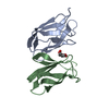
Mass: 92.094 Da / Num. of mol.: 1 / Source method: obtained synthetically / Formula: C3H8O3
#3: Water ChemComp-HOH / water / Water


Mass: 18.015 Da / Num. of mol.: 249 / Source method: isolated from a natural source / Formula: H2O

-
Experimental details

-
Experiment

ExperimentMethod: X-RAY DIFFRACTION / Number of used crystals: 3

-
Sample preparation

CrystalDensity Matthews: 2.22 Å3/Da / Density % sol: 44.67 % / Description: NONE

-
Data collection

DiffractionMean temperature: 100 K
Diffraction sourceSource: SYNCHROTRON / Site: ALBA / Beamline: XALOC / Wavelength: 0.979491
DetectorType: DECTRIS PILATUS 6M / Detector: PIXEL
RadiationProtocol: SINGLE WAVELENGTH / Monochromatic (M) / Laue (L): M / Scattering type: x-ray
Radiation wavelengthWavelength: 0.979491 Å / Relative weight: 1
ReflectionResolution: 0.95→42.73 Å / Num. obs: 92883 / % possible obs: 95.5 % / Observed criterion σ(I): 2 / Redundancy: 2.5 % / Rmerge(I) obs: 0.03 / Net I/σ(I): 13.7
Reflection shellResolution: 0.95→1 Å / Redundancy: 2 % / Rmerge(I) obs: 0.46 / Mean I/σ(I) obs: 1.7 / % possible all: 81.3

-
Processing

Software
NameVersionClassification
REFMAC5.5.0109refinement
XDSdata reduction
CCP4Idata scaling
MOLREPphasing
RefinementMethod to determine structure: MOLECULAR REPLACEMENT / Resolution: 0.95→42.73 Å / Cor.coef. Fo:Fc: 0.974 / Cor.coef. Fo:Fc free: 0.968 / SU B: 0.627 / SU ML: 0.016 / Cross valid method: THROUGHOUT / ESU R: 0.022 / ESU R Free: 0.023 / Stereochemistry target values: MAXIMUM LIKELIHOOD / Details: HYDROGENS HAVE BEEN ADDED IN THE RIDING POSITIONS.
RfactorNum. reflection% reflectionSelection details
Rfree0.17514 4610 5 %RANDOM
Rwork0.15282 ---
obs0.15392 88253 95.47 %-
Solvent computationIon probe radii: 0.8 Å / Shrinkage radii: 0.8 Å / VDW probe radii: 1.4 Å / Solvent model: MASK
Displacement parametersBiso mean: 13.807 Å2
Baniso -1Baniso -2Baniso -3
1-0.07 Å20 Å20.51 Å2
2---0.14 Å20 Å2
3---0.07 Å2
Refinement stepCycle: LAST / Resolution: 0.95→42.73 Å
ProteinNucleic acidLigandSolventTotal
Num. atoms1224 0 6 249 1479
Refine LS restraints
Refine-IDTypeDev idealDev ideal targetNumber
X-RAY DIFFRACTIONr_bond_refined_d0.0290.0221365
X-RAY DIFFRACTIONr_bond_other_d
X-RAY DIFFRACTIONr_angle_refined_deg2.3641.9611871
X-RAY DIFFRACTIONr_angle_other_deg
X-RAY DIFFRACTIONr_dihedral_angle_1_deg6.3765183
X-RAY DIFFRACTIONr_dihedral_angle_2_deg33.18323.250
X-RAY DIFFRACTIONr_dihedral_angle_3_deg13.37615214
X-RAY DIFFRACTIONr_dihedral_angle_4_deg20.856157
X-RAY DIFFRACTIONr_chiral_restr0.4190.2222
X-RAY DIFFRACTIONr_gen_planes_refined0.0150.0211035
X-RAY DIFFRACTIONr_gen_planes_other
X-RAY DIFFRACTIONr_nbd_refined
X-RAY DIFFRACTIONr_nbd_other
X-RAY DIFFRACTIONr_nbtor_refined
X-RAY DIFFRACTIONr_nbtor_other
X-RAY DIFFRACTIONr_xyhbond_nbd_refined
X-RAY DIFFRACTIONr_xyhbond_nbd_other
X-RAY DIFFRACTIONr_metal_ion_refined
X-RAY DIFFRACTIONr_metal_ion_other
X-RAY DIFFRACTIONr_symmetry_vdw_refined
X-RAY DIFFRACTIONr_symmetry_vdw_other
X-RAY DIFFRACTIONr_symmetry_hbond_refined
X-RAY DIFFRACTIONr_symmetry_hbond_other
X-RAY DIFFRACTIONr_symmetry_metal_ion_refined
X-RAY DIFFRACTIONr_symmetry_metal_ion_other
X-RAY DIFFRACTIONr_mcbond_it2.1981.5871
X-RAY DIFFRACTIONr_mcbond_other
X-RAY DIFFRACTIONr_mcangle_it3.21621436
X-RAY DIFFRACTIONr_mcangle_other
X-RAY DIFFRACTIONr_scbond_it4.863494
X-RAY DIFFRACTIONr_scbond_other
X-RAY DIFFRACTIONr_scangle_it6.934.5435
X-RAY DIFFRACTIONr_scangle_other
X-RAY DIFFRACTIONr_long_range_B_refined
X-RAY DIFFRACTIONr_long_range_B_other
X-RAY DIFFRACTIONr_rigid_bond_restr2.59931365
X-RAY DIFFRACTIONr_sphericity_free
X-RAY DIFFRACTIONr_sphericity_bonded
LS refinement shellResolution: 0.949→0.974 Å / Total num. of bins used: 20
RfactorNum. reflection% reflection
Rfree0.324 236 -
Rwork0.313 4862 -
obs--71.36 %
Refinement TLS params.

Method: refined / Refine-ID: X-RAY DIFFRACTION

IDL112)L122)L132)L222)L232)L332)S11 (Å °)S12 (Å °)S13 (Å °)S21 (Å °)S22 (Å °)S23 (Å °)S31 (Å °)S32 (Å °)S33 (Å °)T112)T122)T132)T222)T232)T332)Origin x (Å)Origin y (Å)Origin z (Å)
10.4146-0.0391-0.22050.38360.33870.5276-0.0195-0.0035-0.01540.0096-0.02180.03160.0345-0.00420.04130.0190.0018-0.00140.00450.00280.0133-4.4650.94345.4132
20.78310.2078-0.5010.5318-0.07010.4187-0.0034-0.0408-0.00110.00140.0236-0.0537-0.00240.0372-0.02020.0059-0.0052-0.00180.0178-0.00680.011111.07760.572922.3022
Refinement TLS group
IDRefine-IDRefine TLS-IDAuth asym-IDAuth seq-ID
1X-RAY DIFFRACTION1A383 - 461
2X-RAY DIFFRACTION2B383 - 461

+
About Yorodumi

-
News

-
Feb 9, 2022. New format data for meta-information of EMDB entries

New format data for meta-information of EMDB entries

  • Version 3 of the EMDB header file is now the official format.
  • The previous official version 1.9 will be removed from the archive.

Related info.:EMDB header

External links:wwPDB to switch to version 3 of the EMDB data model

-
Aug 12, 2020. Covid-19 info

Covid-19 info

URL: https://pdbj.org/emnavi/covid19.php

New page: Covid-19 featured information page in EM Navigator.

Related info.:Covid-19 info / Mar 5, 2020. Novel coronavirus structure data

+
Mar 5, 2020. Novel coronavirus structure data

Novel coronavirus structure data

Related info.:Yorodumi Speices / Aug 12, 2020. Covid-19 info

External links:COVID-19 featured content - PDBj / Molecule of the Month (242):Coronavirus Proteases

+
Jan 31, 2019. EMDB accession codes are about to change! (news from PDBe EMDB page)

EMDB accession codes are about to change! (news from PDBe EMDB page)

  • The allocation of 4 digits for EMDB accession codes will soon come to an end. Whilst these codes will remain in use, new EMDB accession codes will include an additional digit and will expand incrementally as the available range of codes is exhausted. The current 4-digit format prefixed with “EMD-” (i.e. EMD-XXXX) will advance to a 5-digit format (i.e. EMD-XXXXX), and so on. It is currently estimated that the 4-digit codes will be depleted around Spring 2019, at which point the 5-digit format will come into force.
  • The EM Navigator/Yorodumi systems omit the EMD- prefix.

Related info.:Q: What is EMD? / ID/Accession-code notation in Yorodumi/EM Navigator

External links:EMDB Accession Codes are Changing Soon! / Contact to PDBj

+
Jul 12, 2017. Major update of PDB

Major update of PDB

  • wwPDB released updated PDB data conforming to the new PDBx/mmCIF dictionary.
  • This is a major update changing the version number from 4 to 5, and with Remediation, in which all the entries are updated.
  • In this update, many items about electron microscopy experimental information are reorganized (e.g. em_software).
  • Now, EM Navigator and Yorodumi are based on the updated data.

External links:wwPDB Remediation / Enriched Model Files Conforming to OneDep Data Standards Now Available in the PDB FTP Archive

-
Yorodumi

Thousand views of thousand structures

  • Yorodumi is a browser for structure data from EMDB, PDB, SASBDB, etc.
  • This page is also the successor to EM Navigator detail page, and also detail information page/front-end page for Omokage search.
  • The word "yorodu" (or yorozu) is an old Japanese word meaning "ten thousand". "mi" (miru) is to see.

Related info.:EMDB / PDB / SASBDB / Comparison of 3 databanks / Yorodumi Search / Aug 31, 2016. New EM Navigator & Yorodumi / Yorodumi Papers / Jmol/JSmol / Function and homology information / Changes in new EM Navigator and Yorodumi

Read more