 Movie
Movie Controller

[English] 日本語
 Yorodumi
- PDB-5ap9: Controlled lid-opening in Thermomyces lanuginosus lipase - a swit... -
+ Open data

- Basic information
| Entry | Database: PDB / ID: 5ap9 | ||||||
|---|---|---|---|---|---|---|---|
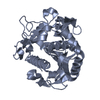
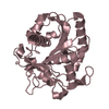
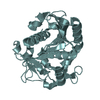
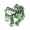
| Title | Controlled lid-opening in Thermomyces lanuginosus lipase - a switch for activity and binding | ||||||
 Components Components | LIPASE | ||||||
 Keywords Keywords | HYDROLASE / THERMOMYCES LANUGINOSUS LIPASE / ENGINEERED DISULFIDE BRIDGE / CONTROLLED BINDING / DUAL SWITCH / CONTROLLED ACTIVITY | ||||||
| Function / homology |  Function and homology informationtriacylglycerol lipase / triglyceride lipase activity / lipid catabolic process Function and homology informationtriacylglycerol lipase / triglyceride lipase activity / lipid catabolic processSimilarity search - Function | ||||||
| Biological species |   THERMOMYCES LANUGINOSUS (fungus) THERMOMYCES LANUGINOSUS (fungus) | ||||||
| Method | X-RAY DIFFRACTION / SYNCHROTRON / MOLECULAR REPLACEMENT / Resolution: 1.8 Å | ||||||
 Authors Authors | Skjold-Joergensen, J. / Vind, J. / Moroz, O.V. / Blagova, E.V. / Bhatia, V.K. / Svendsen, A. / Wilson, K.S. / Bjerrum, M.J. | ||||||
 Citation Citation | Journal: Biochim. Biophys. Acta / Year: 2017 Title: Controlled lid-opening in Thermomyces lanuginosus lipase- An engineered switch for studying lipase function. Authors: Skjold-Jrgensen, J. / Vind, J. / Moroz, O.V. / Blagova, E. / Bhatia, V.K. / Svendsen, A. / Wilson, K.S. / Bjerrum, M.J. | ||||||
| History |
|
- Structure visualization
| Structure viewer | Molecule: MolmilJmol/JSmol |
|---|
- Downloads & links
-Download
| PDBx/mmCIF format | 5ap9.cif.gz | 222.7 KB | Display | PDBx/mmCIF format |
|---|---|---|---|---|
| PDB format | pdb5ap9.ent.gz | 181.2 KB | Display | PDB format |
| PDBx/mmJSON format | 5ap9.json.gz | Tree view | PDBx/mmJSON format | |
| Others |  Other downloads Other downloads |
-Validation report
| Arichive directory | https://data.pdbj.org/pub/pdb/validation_reports/ap/5ap9ftp://data.pdbj.org/pub/pdb/validation_reports/ap/5ap9 | HTTPS FTP |
|---|
-Related structure data
| Related structure data |  1dt3S S: Starting model for refinement |
|---|---|
| Similar structure data |


-Links
 PDBj
PDBj- Assembly
| Deposited unit | 
| ||||||||
|---|---|---|---|---|---|---|---|---|---|
| 1 | 
| ||||||||
| 2 | 
| ||||||||
| Unit cell |
| ||||||||
| Components on special symmetry positions |
|
-Components
-Protein / Sugars , 2 types, 3 molecules AB
| #1: Protein | / TRIACYLGLYCEROL LIPASE Mass: 29322.457 Da / Num. of mol.: 2 / Fragment: UNP RESIDUES 23-291 / Mutation: YES Source method: isolated from a genetically manipulated source Source: (gene. exp.) THERMOMYCES LANUGINOSUS (fungus) / Production host: ASPERGILLUS ORYZAE (mold) / References: UniProt: O59952, triacylglycerol lipase#2: Sugar | ChemComp-NAG / | N-Acetylglucosamine Type: D-saccharide, beta linking / Mass: 221.208 Da / Num. of mol.: 1 Type: D-saccharide, beta linking / Mass: 221.208 Da / Num. of mol.: 1Source method: isolated from a genetically manipulated source Formula: C8H15NO6 |
|---|
-Non-polymers , 4 types, 247 molecules 


| #3: Chemical | Glycerol Mass: 92.094 Da / Num. of mol.: 2 / Source method: obtained synthetically / Formula: C3H8O3 Mass: 92.094 Da / Num. of mol.: 2 / Source method: obtained synthetically / Formula: C3H8O3#4: Chemical | ChemComp-PEG / | Diethylene glycol Mass: 106.120 Da / Num. of mol.: 1 / Source method: obtained synthetically / Formula: C4H10O3 Mass: 106.120 Da / Num. of mol.: 1 / Source method: obtained synthetically / Formula: C4H10O3#5: Chemical | ChemComp-SO4 / | Sulfate Mass: 96.063 Da / Num. of mol.: 1 / Source method: obtained synthetically / Formula: SO4 Mass: 96.063 Da / Num. of mol.: 1 / Source method: obtained synthetically / Formula: SO4#6: Water | ChemComp-HOH / | WaterMass: 18.015 Da / Num. of mol.: 243 / Source method: isolated from a natural source / Formula: H2O |
|---|
-Details
| Sequence details | MUTATIONS I86C, I255C |
|---|
-Experimental details
-Experiment
| Experiment | Method: X-RAY DIFFRACTION / Number of used crystals: 1 |
|---|
- Sample preparation
| Crystal | Density Matthews: 2.8 Å3/Da / Density % sol: 56 % / Description: NONE |
|---|---|
| Crystal grow | Method: vapor diffusion, sitting drop / pH: 4.5 Details: 50 % PEG400, 0.2 M LITHIUM SULFATE, 0.1 M NA-ACETATE PH 4.5, SITTING DROP, VAPOUR DIFFUSION |
-Data collection
| Diffraction | Mean temperature: 110 K |
|---|---|
| Diffraction source | Source: SYNCHROTRON / Site: Diamond  / Beamline: I04 / Wavelength: 0.98 / Beamline: I04 / Wavelength: 0.98 |
| Detector | Date: Jul 25, 2015 |
| Radiation | Protocol: SINGLE WAVELENGTH / Monochromatic (M) / Laue (L): M / Scattering type: x-ray |
| Radiation wavelength | Wavelength: 0.98 Å / Relative weight: 1 |
| Reflection | Resolution: 1.8→50.02 Å / Num. obs: 59391 / % possible obs: 99.9 % / Observed criterion σ(I): 2 / Redundancy: 13.1 % / Rmerge(I) obs: 0.07 / Net I/σ(I): 20.9 |
| Reflection shell | Resolution: 1.8→1.84 Å / Redundancy: 12.8 % / Rmerge(I) obs: 0.87 / Mean I/σ(I) obs: 3 / % possible all: 97.7 |
- Processing
| Software |
| ||||||||||||||||||||||||||||||||||||||||||||||||||||||||||||||||||||||||||||||||||||||||||||||||||||||||||||||||||||||||||||||||||||||||||||||||||||||||||||||||||||||||||||||||||||||
|---|---|---|---|---|---|---|---|---|---|---|---|---|---|---|---|---|---|---|---|---|---|---|---|---|---|---|---|---|---|---|---|---|---|---|---|---|---|---|---|---|---|---|---|---|---|---|---|---|---|---|---|---|---|---|---|---|---|---|---|---|---|---|---|---|---|---|---|---|---|---|---|---|---|---|---|---|---|---|---|---|---|---|---|---|---|---|---|---|---|---|---|---|---|---|---|---|---|---|---|---|---|---|---|---|---|---|---|---|---|---|---|---|---|---|---|---|---|---|---|---|---|---|---|---|---|---|---|---|---|---|---|---|---|---|---|---|---|---|---|---|---|---|---|---|---|---|---|---|---|---|---|---|---|---|---|---|---|---|---|---|---|---|---|---|---|---|---|---|---|---|---|---|---|---|---|---|---|---|---|---|---|---|---|
| Refinement | Method to determine structure: MOLECULAR REPLACEMENT Starting model: PDB ENTRY 1DT3 Resolution: 1.8→50.02 Å / Cor.coef. Fo:Fc: 0.965 / Cor.coef. Fo:Fc free: 0.951 / SU B: 4.243 / SU ML: 0.067 / Cross valid method: THROUGHOUT / ESU R: 0.102 / ESU R Free: 0.098 / Stereochemistry target values: MAXIMUM LIKELIHOOD Details: HYDROGENS HAVE BEEN ADDED IN THE RIDING POSITIONS. SUBUNIT A IS BETTER ORDERED, THAN SUBUNIT B. DESCRIPTION IN THE PAPER WILL REFER TO SUBUNIT A
| ||||||||||||||||||||||||||||||||||||||||||||||||||||||||||||||||||||||||||||||||||||||||||||||||||||||||||||||||||||||||||||||||||||||||||||||||||||||||||||||||||||||||||||||||||||||
| Solvent computation | Ion probe radii: 0.8 Å / Shrinkage radii: 0.8 Å / VDW probe radii: 1.2 Å / Solvent model: MASK | ||||||||||||||||||||||||||||||||||||||||||||||||||||||||||||||||||||||||||||||||||||||||||||||||||||||||||||||||||||||||||||||||||||||||||||||||||||||||||||||||||||||||||||||||||||||
| Displacement parameters | Biso mean: 36.267 Å2
| ||||||||||||||||||||||||||||||||||||||||||||||||||||||||||||||||||||||||||||||||||||||||||||||||||||||||||||||||||||||||||||||||||||||||||||||||||||||||||||||||||||||||||||||||||||||
| Refinement step | Cycle: LAST / Resolution: 1.8→50.02 Å
| ||||||||||||||||||||||||||||||||||||||||||||||||||||||||||||||||||||||||||||||||||||||||||||||||||||||||||||||||||||||||||||||||||||||||||||||||||||||||||||||||||||||||||||||||||||||
| Refine LS restraints |
|