 Movie
Movie Controller


+ Open data

- Basic information
| Entry | Database: PDB / ID: 5an5 | |||||||||
|---|---|---|---|---|---|---|---|---|---|---|
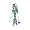
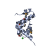
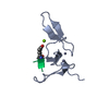
| Title | B. subtilis GpsB C-terminal Domain | |||||||||
 Components Components | CELL CYCLE PROTEIN GPSB | |||||||||
 Keywords Keywords | CELL CYCLE / BACTERIAL GROWTH REGULATION / CELL WALL SYNTHESIS / CELL DIVISION | |||||||||
| Function / homology | Cell cycle protein GpsB / DivIVA family / DivIVA domain / DivIVA protein / regulation of cell shape / cell cycle / cell division / cytoplasm / Cell cycle protein GpsB Function and homology information Function and homology information | |||||||||
| Biological species |  BACILLUS SUBTILIS SUBSP. SUBTILIS STR. 168 (bacteria) BACILLUS SUBTILIS SUBSP. SUBTILIS STR. 168 (bacteria) | |||||||||
| Method | X-RAY DIFFRACTION / SYNCHROTRON / OTHER / Resolution: 1.2 Å | |||||||||
 Authors Authors | Rismondo, J. / Cleverley, R.M. / Lane, H.V. / Grohennig, S. / Steglich, A. / Moller, L. / Krishna Mannala, G. / Hain, T. / Lewis, R.J. / Halbedel, S. | |||||||||
 Citation Citation | Journal: Mol.Microbiol. / Year: 2016 Title: Structure of the Bacterial Cell Division Determinant Gpsb and its Interaction with Penicillin Binding Proteins. Authors: Rismondo, J. / Cleverley, R.M. / Lane, H.V. / Grosshennig, S. / Steglich, A. / Moller, L. / Mannala, G.K. / Hain, T. / Lewis, R.J. / Halbedel, S. | |||||||||
| History |
|
- Structure visualization
| Structure viewer | Molecule: MolmilJmol/JSmol |
|---|
- Downloads & links
-Download
| PDBx/mmCIF format | 5an5.cif.gz | 89.1 KB | Display | PDBx/mmCIF format |
|---|---|---|---|---|
| PDB format | pdb5an5.ent.gz | 71.9 KB | Display | PDB format |
| PDBx/mmJSON format | 5an5.json.gz | Tree view | PDBx/mmJSON format | |
| Others |  Other downloads Other downloads |
-Validation report
| Arichive directory | https://data.pdbj.org/pub/pdb/validation_reports/an/5an5ftp://data.pdbj.org/pub/pdb/validation_reports/an/5an5 | HTTPS FTP |
|---|
-Related structure data


-Links
 PDBj
PDBj- Assembly
| Deposited unit | 
| ||||||||
|---|---|---|---|---|---|---|---|---|---|
| 1 | 
| ||||||||
| 2 | 
| ||||||||
| 3 | 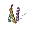
| ||||||||
| Unit cell |
|
-Components
| #1: Protein/peptide | / GUIDING PBP1-SHUTTLING PROTEIN / GPSB Mass: 3089.546 Da / Num. of mol.: 9 / Fragment: C-TERMINAL DOMAIN, UNP RESIDUES 76-98 Source method: isolated from a genetically manipulated source Source: (gene. exp.) BACILLUS SUBTILIS SUBSP. SUBTILIS STR. 168 (bacteria)Production host: ESCHERICHIA COLI (E. coli) / Strain (production host): BL21(DE3) / References: UniProt: P0CI74#2: Chemical | Glycerol  Mass: 92.094 Da / Num. of mol.: 2 / Source method: obtained synthetically / Formula: C3H8O3 Mass: 92.094 Da / Num. of mol.: 2 / Source method: obtained synthetically / Formula: C3H8O3#3: Water | ChemComp-HOH / | Water Mass: 18.015 Da / Num. of mol.: 235 / Source method: isolated from a natural source / Formula: H2O Mass: 18.015 Da / Num. of mol.: 235 / Source method: isolated from a natural source / Formula: H2OSequence details | RESIDUES NUMBERED -4 TO 0 (GMSA SEQUENCE) ARE DERIVED FROM THE EXPRESSION | |
|---|
-Experimental details
-Experiment
| Experiment | Method: X-RAY DIFFRACTION / Number of used crystals: 1 |
|---|
- Sample preparation
| Crystal | Density Matthews: 1.67 Å3/Da / Density % sol: 26.19 % / Description: NONE |
|---|---|
| Crystal grow | pH: 7.5 / Details: 0.2M SODIUM FLUORIDE, 20% PEG 3350, pH 7.5 |
-Data collection
| Diffraction | Mean temperature: 100 K |
|---|---|
| Diffraction source | Source: SYNCHROTRON / Site: Diamond  / Beamline: I03 / Wavelength: 0.61992 / Beamline: I03 / Wavelength: 0.61992 |
| Detector | Type: DECTRIS PILATUS 6M / Detector: PIXEL / Date: Jul 4, 2015 |
| Radiation | Protocol: SINGLE WAVELENGTH / Monochromatic (M) / Laue (L): M / Scattering type: x-ray |
| Radiation wavelength | Wavelength: 0.61992 Å / Relative weight: 1 |
| Reflection | Resolution: 1.2→43.04 Å / Num. obs: 54744 / % possible obs: 97.8 % / Observed criterion σ(I): 0 / Redundancy: 5.3 % / Biso Wilson estimate: 10.33 Å2 / Rmerge(I) obs: 0.05 / Net I/σ(I): 57.4 |
| Reflection shell | Resolution: 1.2→1.22 Å / Redundancy: 5.1 % / Rmerge(I) obs: 0.13 / Mean I/σ(I) obs: 14.2 / % possible all: 96.6 |
- Processing
| Software |
| |||||||||||||||||||||||||||||||||||||||||||||||||||||||||||||||
|---|---|---|---|---|---|---|---|---|---|---|---|---|---|---|---|---|---|---|---|---|---|---|---|---|---|---|---|---|---|---|---|---|---|---|---|---|---|---|---|---|---|---|---|---|---|---|---|---|---|---|---|---|---|---|---|---|---|---|---|---|---|---|---|---|
| Refinement | Method to determine structure: OTHER Starting model: NONE Resolution: 1.2→29.659 Å / SU ML: 0.08 / σ(F): 1.98 / Phase error: 16.53 / Stereochemistry target values: ML
| |||||||||||||||||||||||||||||||||||||||||||||||||||||||||||||||
| Solvent computation | Shrinkage radii: 0.9 Å / VDW probe radii: 1.11 Å / Solvent model: FLAT BULK SOLVENT MODEL / Bsol: 58.6 Å2 / ksol: 0.43 e/Å3 | |||||||||||||||||||||||||||||||||||||||||||||||||||||||||||||||
| Displacement parameters | Biso mean: 16 Å2 | |||||||||||||||||||||||||||||||||||||||||||||||||||||||||||||||
| Refinement step | Cycle: LAST / Resolution: 1.2→29.659 Å
| |||||||||||||||||||||||||||||||||||||||||||||||||||||||||||||||
| Refine LS restraints |
| |||||||||||||||||||||||||||||||||||||||||||||||||||||||||||||||
| LS refinement shell |
|
