 Movie
Movie Controller

[English] 日本語
 Yorodumi
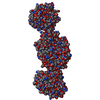
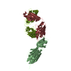
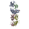
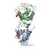
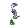
- PDB-5a6s: Crystal structure of the CTP1L endolysin reveals how its activity... -
+ Open data

- Basic information
| Entry | Database: PDB / ID: 5a6s | ||||||
|---|---|---|---|---|---|---|---|
| Title | Crystal structure of the CTP1L endolysin reveals how its activity is regulated by a secondary translation product | ||||||
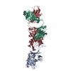
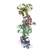
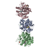
 Components Components | (ENDOLYSIN Lysin) x 2 Lysin) x 2 | ||||||
 Keywords Keywords | STRUCTURAL PROTEIN / ENDOLYSIN / SECONDARY TRANSLATION PRODUCT / BACTERIOPHAGE | ||||||
| Function / homology |  Function and homology information Function and homology informationpeptidoglycan catabolic process / cell wall macromolecule catabolic process / lysozyme / lysozyme activitySimilarity search - Function | ||||||
| Biological species |  CLOSTRIDIUM PHAGE PHICTP1 (virus) CLOSTRIDIUM PHAGE PHICTP1 (virus) | ||||||
| Method | X-RAY DIFFRACTION / SYNCHROTRON / MOLECULAR REPLACEMENT / Resolution: 1.9 Å | ||||||
 Authors Authors | Dunne, M. / Leicht, S. / Krichel, B. / Mertens, H.D.T. / Thompson, A. / Krijgsveld, J. / Svergun, D.I. / GomezTorres, N. / Garde, S. / Uetrecht, C. ...Dunne, M. / Leicht, S. / Krichel, B. / Mertens, H.D.T. / Thompson, A. / Krijgsveld, J. / Svergun, D.I. / GomezTorres, N. / Garde, S. / Uetrecht, C. / Narbad, A. / Mayer, M.J. / Meijers, R. | ||||||
 Citation Citation | Journal: J Biol Chem / Year: 2016 Title: Crystal Structure of the CTP1L Endolysin Reveals How Its Activity Is Regulated by a Secondary Translation Product. Authors: Matthew Dunne / Stefan Leicht / Boris Krichel / Haydyn D T Mertens / Andrew Thompson / Jeroen Krijgsveld / Dmitri I Svergun / Natalia Gómez-Torres / Sonia Garde / Charlotte Uetrecht / Arjan ...Authors: Matthew Dunne / Stefan Leicht / Boris Krichel / Haydyn D T Mertens / Andrew Thompson / Jeroen Krijgsveld / Dmitri I Svergun / Natalia Gómez-Torres / Sonia Garde / Charlotte Uetrecht / Arjan Narbad / Melinda J Mayer / Rob Meijers /     Abstract: Bacteriophages produce endolysins, which lyse the bacterial host cell to release newly produced virions. The timing of lysis is regulated and is thought to involve the activation of a molecular ...Bacteriophages produce endolysins, which lyse the bacterial host cell to release newly produced virions. The timing of lysis is regulated and is thought to involve the activation of a molecular switch. We present a crystal structure of the activated endolysin CTP1L that targets Clostridium tyrobutyricum, consisting of a complex between the full-length protein and an N-terminally truncated C-terminal cell wall binding domain (CBD). The truncated CBD is produced through an internal translation start site within the endolysin gene. Mutants affecting the internal translation site change the oligomeric state of the endolysin and reduce lytic activity. The activity can be modulated by reconstitution of the full-length endolysin-CBD complex with free CBD. The same oligomerization mechanism applies to the CD27L endolysin that targets Clostridium difficile and the CS74L endolysin that targets Clostridium sporogenes. When the CTP1L endolysin gene is introduced into the commensal bacterium Lactococcus lactis, the truncated CBD is also produced, showing that the alternative start codon can be used in other bacterial species. The identification of a translational switch affecting oligomerization presented here has implications for the design of effective endolysins for the treatment of bacterial infections. | ||||||
| History |
| ||||||
| Remark 700 | SHEET DETERMINATION METHOD: DSSP THE SHEETS PRESENTED AS "AA" IN EACH CHAIN ON SHEET RECORDS BELOW ... SHEET DETERMINATION METHOD: DSSP THE SHEETS PRESENTED AS "AA" IN EACH CHAIN ON SHEET RECORDS BELOW IS ACTUALLY AN 8-STRANDED BARREL THIS IS REPRESENTED BY A 9-STRANDED SHEET IN WHICH THE FIRST AND LAST STRANDS ARE IDENTICAL. |
- Structure visualization
| Structure viewer | Molecule: MolmilJmol/JSmol |
|---|
- Downloads & links
-Download
| PDBx/mmCIF format | 5a6s.cif.gz | 95.8 KB | Display | PDBx/mmCIF format |
|---|---|---|---|---|
| PDB format | pdb5a6s.ent.gz | 72 KB | Display | PDB format |
| PDBx/mmJSON format | 5a6s.json.gz | Tree view | PDBx/mmJSON format | |
| Others |  Other downloads Other downloads |
-Validation report
| Arichive directory | https://data.pdbj.org/pub/pdb/validation_reports/a6/5a6sftp://data.pdbj.org/pub/pdb/validation_reports/a6/5a6s | HTTPS FTP |
|---|
-Related structure data
| Related structure data |  1jfxS  2nw0S S: Starting model for refinement C: citing same article ( |
|---|---|
| Similar structure data |


-Links
 PDBj
PDBj- Assembly
| Deposited unit | 
| ||||||||
|---|---|---|---|---|---|---|---|---|---|
| 1 | 
| ||||||||
| Unit cell |
|
-Components
-Protein , 2 types, 2 molecules AB
| #1: Protein | Lysin Mass: 32877.180 Da / Num. of mol.: 1 Source method: isolated from a genetically manipulated source Source: (gene. exp.) CLOSTRIDIUM PHAGE PHICTP1 (virus) / Strain: CTP1L / Description: TARGET AGAINST CLOSTRIDIUM TYROBUTYRICUM / Production host:  ESCHERICHIA COLI (E. coli) / Strain (production host): BL21(DE3) / References: UniProt: D9ZNF3 ESCHERICHIA COLI (E. coli) / Strain (production host): BL21(DE3) / References: UniProt: D9ZNF3 |
|---|---|
| #2: Protein | Lysin Mass: 9031.121 Da / Num. of mol.: 1 Fragment: TRUNCATED CELL WALL BINDING DOMAIN, RESIDUES 195-274 Mutation: YES Source method: isolated from a genetically manipulated source Source: (gene. exp.) CLOSTRIDIUM PHAGE PHICTP1 (virus) / Production host: ESCHERICHIA COLI (E. coli) / Strain (production host): BL21(DE3) / References: UniProt: D9ZNF3 |
-Non-polymers , 4 types, 485 molecules 


| #3: Chemical | ChemComp-SO4 / Sulfate Mass: 96.063 Da / Num. of mol.: 1 / Source method: obtained synthetically / Formula: SO4 Mass: 96.063 Da / Num. of mol.: 1 / Source method: obtained synthetically / Formula: SO4 | ||||
|---|---|---|---|---|---|
| #4: Chemical | Glycerol Mass: 92.094 Da / Num. of mol.: 3 / Source method: obtained synthetically / Formula: C3H8O3 Mass: 92.094 Da / Num. of mol.: 3 / Source method: obtained synthetically / Formula: C3H8O3#5: Chemical | ChemComp-1PE / | Polyethylene glycol Mass: 238.278 Da / Num. of mol.: 1 / Source method: obtained synthetically / Formula: C10H22O6 / Comment: precipitant*YM Mass: 238.278 Da / Num. of mol.: 1 / Source method: obtained synthetically / Formula: C10H22O6 / Comment: precipitant*YM#6: Water | ChemComp-HOH / | WaterMass: 18.015 Da / Num. of mol.: 480 / Source method: isolated from a natural source / Formula: H2O |
-Details
| Sequence details | THIS CHAIN IS A TRUNCATED CELL WALL BINDING DOMAIN THAT IS THE RESULT OF SECONDARY TRANSLATION ...THIS CHAIN IS A TRUNCATED CELL WALL BINDING DOMAIN THAT IS THE RESULT OF SECONDARY TRANSLATIO |
|---|
-Experimental details
-Experiment
| Experiment | Method: X-RAY DIFFRACTION / Number of used crystals: 1 |
|---|
- Sample preparation
| Crystal | Density Matthews: 3.12 Å3/Da / Density % sol: 60.62 % / Description: NONE |
|---|---|
| Crystal grow | Details: 5-10 % PEG 8000, 20 MM TRIS PH 8.0 |
-Data collection
| Diffraction | Mean temperature: 100 K |
|---|---|
| Diffraction source | Source: SYNCHROTRON / Site: SOLEIL / Beamline: PROXIMA 1 / Wavelength: 0.98 |
| Detector | Type: ADSC CCD / Detector: CCD / Details: KB MIRRORS |
| Radiation | Monochromator: SI 1 1 1 / Protocol: SINGLE WAVELENGTH / Monochromatic (M) / Laue (L): M / Scattering type: x-ray |
| Radiation wavelength | Wavelength: 0.98 Å / Relative weight: 1 |
| Reflection | Resolution: 1.9→30 Å / Num. obs: 40146 / % possible obs: 100 % / Observed criterion σ(I): 0 / Redundancy: 8.2 % / Rmerge(I) obs: 0.15 / Net I/σ(I): 9.6 |
| Reflection shell | Resolution: 1.9→2 Å / Redundancy: 8 % / Rmerge(I) obs: 0.75 / Mean I/σ(I) obs: 2.6 / % possible all: 100 |
- Processing
| Software |
| ||||||||||||||||||||||||||||||||||||||||||||||||||||||||||||||||||||||||||||||||||||||||||||||||||||||||||||||||||||||||||||||||||||||||||||||||||||||||||||||||||||||||||||||||||||||
|---|---|---|---|---|---|---|---|---|---|---|---|---|---|---|---|---|---|---|---|---|---|---|---|---|---|---|---|---|---|---|---|---|---|---|---|---|---|---|---|---|---|---|---|---|---|---|---|---|---|---|---|---|---|---|---|---|---|---|---|---|---|---|---|---|---|---|---|---|---|---|---|---|---|---|---|---|---|---|---|---|---|---|---|---|---|---|---|---|---|---|---|---|---|---|---|---|---|---|---|---|---|---|---|---|---|---|---|---|---|---|---|---|---|---|---|---|---|---|---|---|---|---|---|---|---|---|---|---|---|---|---|---|---|---|---|---|---|---|---|---|---|---|---|---|---|---|---|---|---|---|---|---|---|---|---|---|---|---|---|---|---|---|---|---|---|---|---|---|---|---|---|---|---|---|---|---|---|---|---|---|---|---|---|
| Refinement | Method to determine structure: MOLECULAR REPLACEMENT Starting model: PDB ENTRIES 1JFX AND 2NW0 HYBRID MODEL Resolution: 1.9→19.9 Å / Cor.coef. Fo:Fc: 0.963 / Cor.coef. Fo:Fc free: 0.943 / SU B: 2.55 / SU ML: 0.075 / Cross valid method: THROUGHOUT / ESU R: 0.11 / ESU R Free: 0.114 / Stereochemistry target values: MAXIMUM LIKELIHOOD / Details: HYDROGENS HAVE BEEN ADDED IN THE RIDING POSITIONS.
| ||||||||||||||||||||||||||||||||||||||||||||||||||||||||||||||||||||||||||||||||||||||||||||||||||||||||||||||||||||||||||||||||||||||||||||||||||||||||||||||||||||||||||||||||||||||
| Solvent computation | Ion probe radii: 0.8 Å / Shrinkage radii: 0.8 Å / VDW probe radii: 1.2 Å / Solvent model: MASK | ||||||||||||||||||||||||||||||||||||||||||||||||||||||||||||||||||||||||||||||||||||||||||||||||||||||||||||||||||||||||||||||||||||||||||||||||||||||||||||||||||||||||||||||||||||||
| Displacement parameters | Biso mean: 26.486 Å2
| ||||||||||||||||||||||||||||||||||||||||||||||||||||||||||||||||||||||||||||||||||||||||||||||||||||||||||||||||||||||||||||||||||||||||||||||||||||||||||||||||||||||||||||||||||||||
| Refinement step | Cycle: LAST / Resolution: 1.9→19.9 Å
| ||||||||||||||||||||||||||||||||||||||||||||||||||||||||||||||||||||||||||||||||||||||||||||||||||||||||||||||||||||||||||||||||||||||||||||||||||||||||||||||||||||||||||||||||||||||
| Refine LS restraints |
|