 Movie
Movie Controller

[English] 日本語
 Yorodumi
- PDB-5a6l: High resolution structure of the thermostable glucuronoxylan endo... -
+ Open data

- Basic information
| Entry | Database: PDB / ID: 5a6l | |||||||||
|---|---|---|---|---|---|---|---|---|---|---|
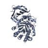
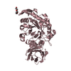
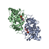
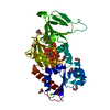
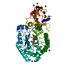
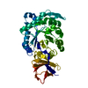
| Title | High resolution structure of the thermostable glucuronoxylan endo-Beta-1, 4-xylanase, CtXyn30A, from Clostridium thermocellum with two xylobiose units bound | |||||||||
 Components Components | CARBOHYDRATE BINDING FAMILY 6 | |||||||||
 Keywords Keywords |  HYDROLASE / PROTEIN HYDROLASE / PROTEIN | |||||||||
| Function / homology |  Function and homology informationglucosylceramidase activity / sphingolipid metabolic process / polysaccharide catabolic process / carbohydrate binding Function and homology informationglucosylceramidase activity / sphingolipid metabolic process / polysaccharide catabolic process / carbohydrate bindingSimilarity search - Function | |||||||||
| Biological species |  CLOSTRIDIUM THERMOCELLUM (bacteria) CLOSTRIDIUM THERMOCELLUM (bacteria) | |||||||||
| Method | X-RAY DIFFRACTION / SYNCHROTRON / MOLECULAR REPLACEMENT / Resolution: 1.8 Å | |||||||||
 Authors Authors | Freire, F. / Verma, A.K. / Bule, P. / Goyal, A. / Fontes, C.M.G.A. / Najmudin, S. | |||||||||
 Citation Citation | Journal: Acta Crystallogr.,Sect.D / Year: 2016 Title: Conservation in the Mechanism of Glucuronoxylan Hydrolysis Revealed by the Structure of Glucuronoxylan Xylano-Hydrolase (Ctxyn30A) from Clostridium Thermocellum Authors: Freire, F. / Verma, A.K. / Bule, P. / Alves, V.D. / Fontes, C.M.G.A. / Goyal, A. / Najmudin, S. | |||||||||
| History |
| |||||||||
| Remark 700 | SHEET DETERMINATION METHOD: DSSP THE SHEETS PRESENTED AS "AB" IN EACH CHAIN ON SHEET RECORDS BELOW ... SHEET DETERMINATION METHOD: DSSP THE SHEETS PRESENTED AS "AB" IN EACH CHAIN ON SHEET RECORDS BELOW IS ACTUALLY AN 8-STRANDED BARREL THIS IS REPRESENTED BY A 9-STRANDED SHEET IN WHICH THE FIRST AND LAST STRANDS ARE IDENTICAL. |
- Structure visualization
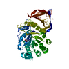
| Structure viewer | Molecule: MolmilJmol/JSmol |
|---|
- Downloads & links
-Download
| PDBx/mmCIF format | 5a6l.cif.gz | 175.2 KB | Display | PDBx/mmCIF format |
|---|---|---|---|---|
| PDB format | pdb5a6l.ent.gz | 137.7 KB | Display | PDB format |
| PDBx/mmJSON format | 5a6l.json.gz | Tree view | PDBx/mmJSON format | |
| Others |  Other downloads Other downloads |
-Validation report
| Arichive directory | https://data.pdbj.org/pub/pdb/validation_reports/a6/5a6lftp://data.pdbj.org/pub/pdb/validation_reports/a6/5a6l | HTTPS FTP |
|---|
-Related structure data
| Related structure data |  4ckqC 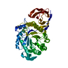 4uq9C  4uqaC 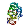 4uqbC 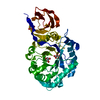 4uqcC 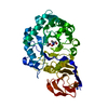 4uqdC  4uqeSC 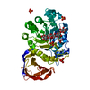 5a6mC C: citing same article ( S: Starting model for refinement |
|---|---|
| Similar structure data |


-Links
 PDBj
PDBj- Assembly
| Deposited unit | 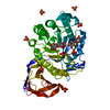
| ||||||||
|---|---|---|---|---|---|---|---|---|---|
| 1 |
| ||||||||
| Unit cell |
|
-Components
| #1: Protein | Mass: 46396.027 Da / Num. of mol.: 1 / Fragment: N-TERMINAL CATALYTIC MODULE, UNP RESIDUES 34-419 / Mutation: YES Source method: isolated from a genetically manipulated source Source: (gene. exp.) CLOSTRIDIUM THERMOCELLUM (bacteria) / Plasmid: PET28A / Production host: ESCHERICHIA COLI (E. coli) / Strain (production host): BL21(DE3) / References: UniProt: A3DJS9, endo-1,4-beta-xylanase | ||||||
|---|---|---|---|---|---|---|---|
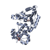
| #2: Polysaccharide | 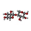  , Oligosaccharide / Class: Metabolism / Mass: 282.245 Da / Num. of mol.: 2 , Oligosaccharide / Class: Metabolism / Mass: 282.245 Da / Num. of mol.: 2Source method: isolated from a genetically manipulated source Details: oligosaccharide / References: 4beta-beta-xylobiose #3: Chemical | ChemComp-PO4 / Phosphate  Mass: 94.971 Da / Num. of mol.: 8 / Source method: obtained synthetically / Formula: PO4 Mass: 94.971 Da / Num. of mol.: 8 / Source method: obtained synthetically / Formula: PO4#4: Chemical | ChemComp-PEG / | Diethylene glycol  Mass: 106.120 Da / Num. of mol.: 1 / Source method: obtained synthetically / Formula: C4H10O3 Mass: 106.120 Da / Num. of mol.: 1 / Source method: obtained synthetically / Formula: C4H10O3#5: Water | ChemComp-HOH / | Water Mass: 18.015 Da / Num. of mol.: 404 / Source method: isolated from a natural source / Formula: H2O Mass: 18.015 Da / Num. of mol.: 404 / Source method: isolated from a natural source / Formula: H2O |
-Experimental details
-Experiment
| Experiment | Method: X-RAY DIFFRACTION / Number of used crystals: 1 |
|---|
- Sample preparation
| Crystal | Density Matthews: 2.57 Å3/Da / Density % sol: 52.25 % / Description: NONE |
|---|---|
| Crystal grow | Details: 0.2 M POTASSIUM PHOSPHATE |
-Data collection
| Diffraction | Mean temperature: 100 K |
|---|---|
| Diffraction source | Source: SYNCHROTRON / Site: SOLEIL  / Beamline: PROXIMA 1 / Wavelength: 0.95373 / Beamline: PROXIMA 1 / Wavelength: 0.95373 |
| Detector | Type: DECTRIS PILATUS 6M / Detector: PIXEL / Date: Dec 17, 2014 |
| Radiation | Monochromator: CHANNEL CUT CRYOGENICALLY COOLED MONOCHROMATOR CRYSTAL Protocol: SINGLE WAVELENGTH / Monochromatic (M) / Laue (L): M / Scattering type: x-ray |
| Radiation wavelength | Wavelength: 0.95373 Å / Relative weight: 1 |
| Reflection | Resolution: 1.8→46.25 Å / Num. obs: 43517 / % possible obs: 100 % / Observed criterion σ(I): 9.8 / Redundancy: 3.8 % / Rmerge(I) obs: 0.28 / Net I/σ(I): 17.5 |
| Reflection shell | Resolution: 1.8→1.84 Å / Redundancy: 3.8 % / Rmerge(I) obs: 1.49 / Mean I/σ(I) obs: 9.8 / % possible all: 100 |
- Processing
| Software |
| ||||||||||||||||||||||||||||||||||||||||||||||||||||||||||||||||||||||||||||||||||||||||||||||||||||||||||||||||||||||||||||||||||||||||||||||||||||||||||||||||||||||||||||||||||||||
|---|---|---|---|---|---|---|---|---|---|---|---|---|---|---|---|---|---|---|---|---|---|---|---|---|---|---|---|---|---|---|---|---|---|---|---|---|---|---|---|---|---|---|---|---|---|---|---|---|---|---|---|---|---|---|---|---|---|---|---|---|---|---|---|---|---|---|---|---|---|---|---|---|---|---|---|---|---|---|---|---|---|---|---|---|---|---|---|---|---|---|---|---|---|---|---|---|---|---|---|---|---|---|---|---|---|---|---|---|---|---|---|---|---|---|---|---|---|---|---|---|---|---|---|---|---|---|---|---|---|---|---|---|---|---|---|---|---|---|---|---|---|---|---|---|---|---|---|---|---|---|---|---|---|---|---|---|---|---|---|---|---|---|---|---|---|---|---|---|---|---|---|---|---|---|---|---|---|---|---|---|---|---|---|
| Refinement | Method to determine structure: MOLECULAR REPLACEMENT Starting model: PDBE ENTRY 4UQE Resolution: 1.8→54.54 Å / Cor.coef. Fo:Fc: 0.964 / Cor.coef. Fo:Fc free: 0.95 / SU B: 3.268 / SU ML: 0.054 / Cross valid method: THROUGHOUT / ESU R: 0.092 / ESU R Free: 0.089 / Stereochemistry target values: MAXIMUM LIKELIHOOD Details: HYDROGENS HAVE BEEN ADDED IN THE RIDING POSITIONS. U VALUES WITH TLS ADDED
| ||||||||||||||||||||||||||||||||||||||||||||||||||||||||||||||||||||||||||||||||||||||||||||||||||||||||||||||||||||||||||||||||||||||||||||||||||||||||||||||||||||||||||||||||||||||
| Solvent computation | Ion probe radii: 0.7 Å / Shrinkage radii: 0.7 Å / VDW probe radii: 1.3 Å / Solvent model: MASK | ||||||||||||||||||||||||||||||||||||||||||||||||||||||||||||||||||||||||||||||||||||||||||||||||||||||||||||||||||||||||||||||||||||||||||||||||||||||||||||||||||||||||||||||||||||||
| Displacement parameters | Biso mean: 9.888 Å2
| ||||||||||||||||||||||||||||||||||||||||||||||||||||||||||||||||||||||||||||||||||||||||||||||||||||||||||||||||||||||||||||||||||||||||||||||||||||||||||||||||||||||||||||||||||||||
| Refinement step | Cycle: LAST / Resolution: 1.8→54.54 Å
| ||||||||||||||||||||||||||||||||||||||||||||||||||||||||||||||||||||||||||||||||||||||||||||||||||||||||||||||||||||||||||||||||||||||||||||||||||||||||||||||||||||||||||||||||||||||
| Refine LS restraints |
|